Nikon Small World announced the winners of its 2010 photomicrography contest.
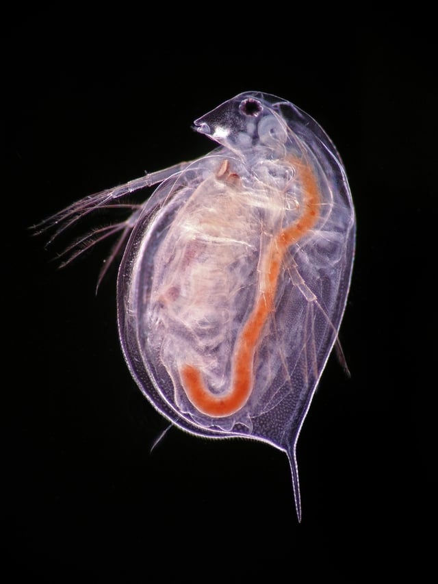
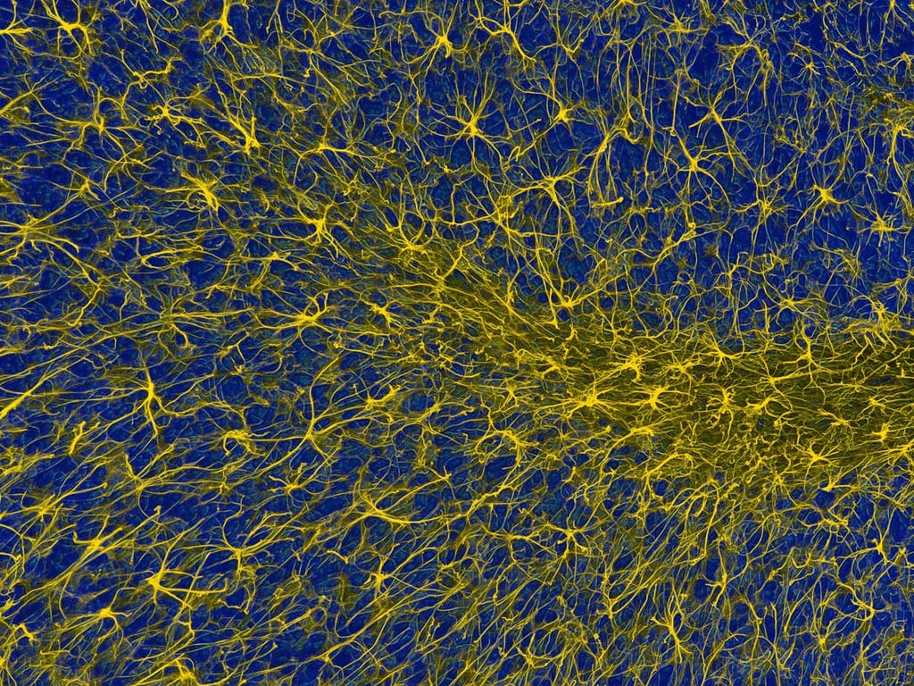
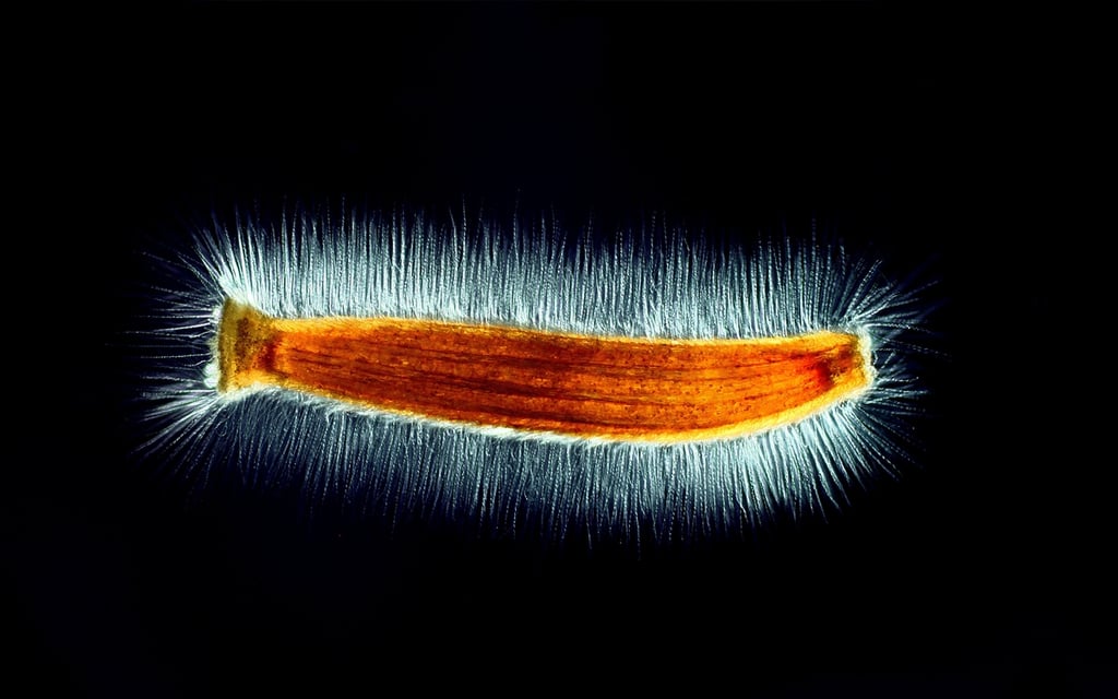
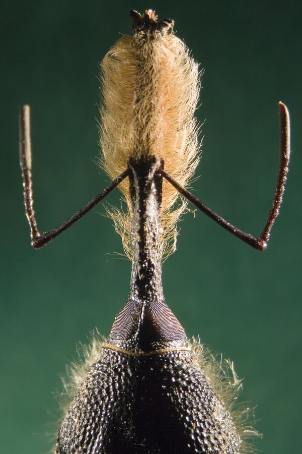
The winning photo was taken by Jonas King – Vanderbilt University, Department of Biological Sciences in Nashville, Tennessee
Courtesy of Nikon Small World
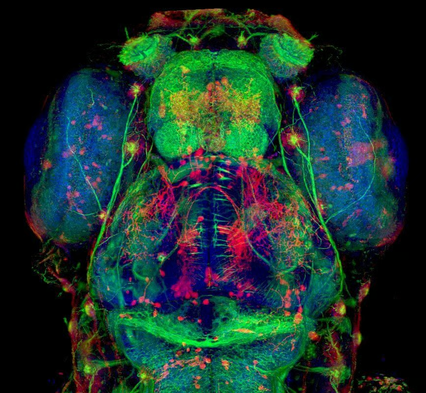
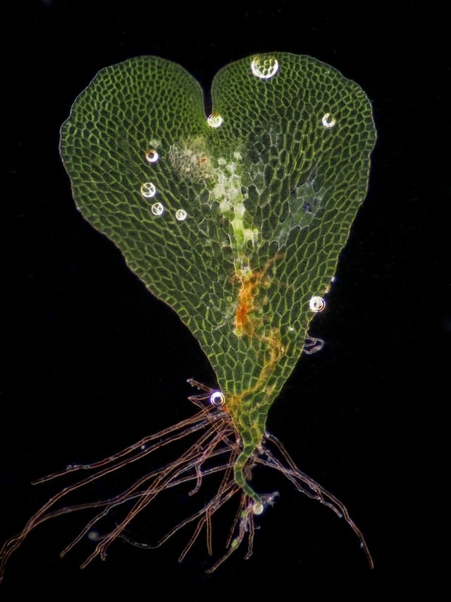
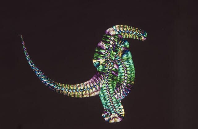
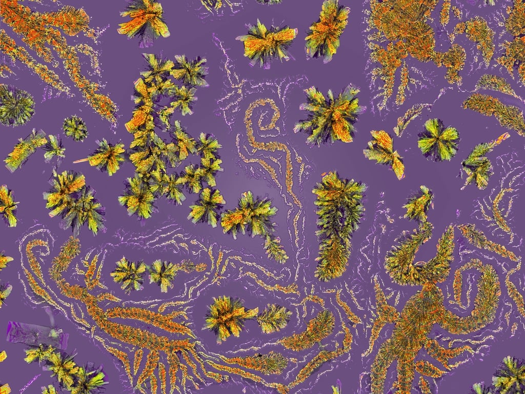
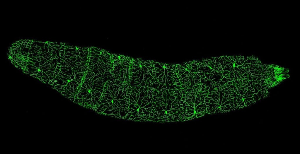
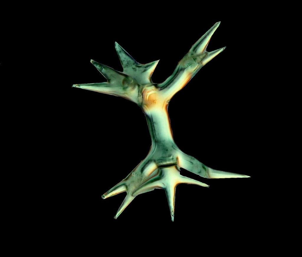
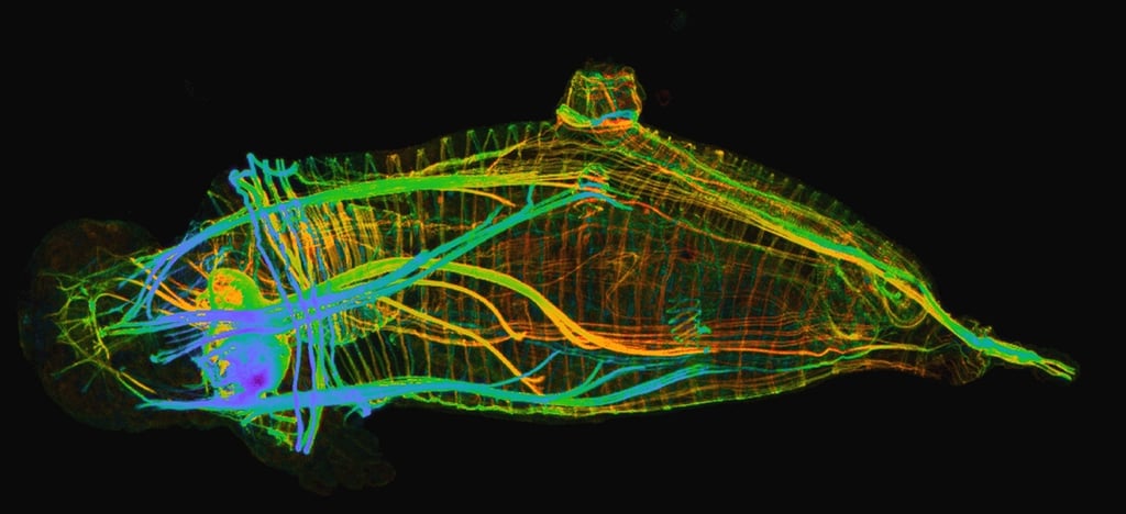
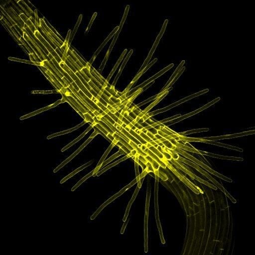
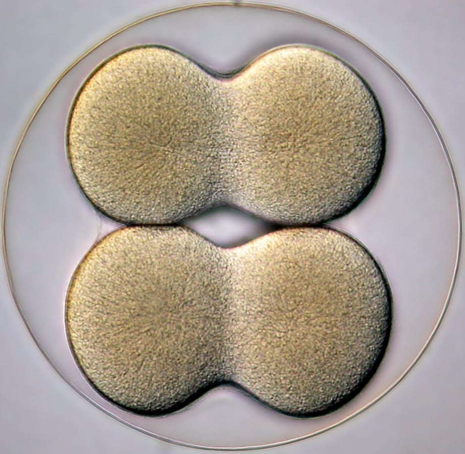
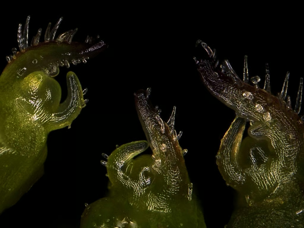
Dr. Hideo Otsuna – University of Utah Medical Center, Department of Neurobiology and Anatomy in Salt Lake City, Utah
Courtesy of Nikon Small World
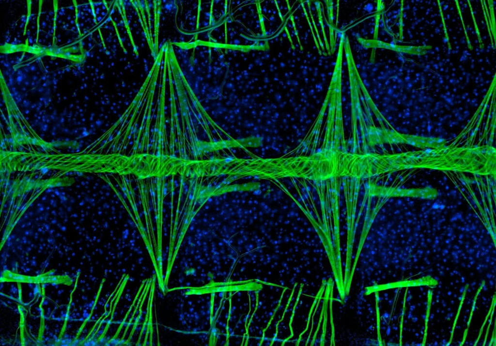
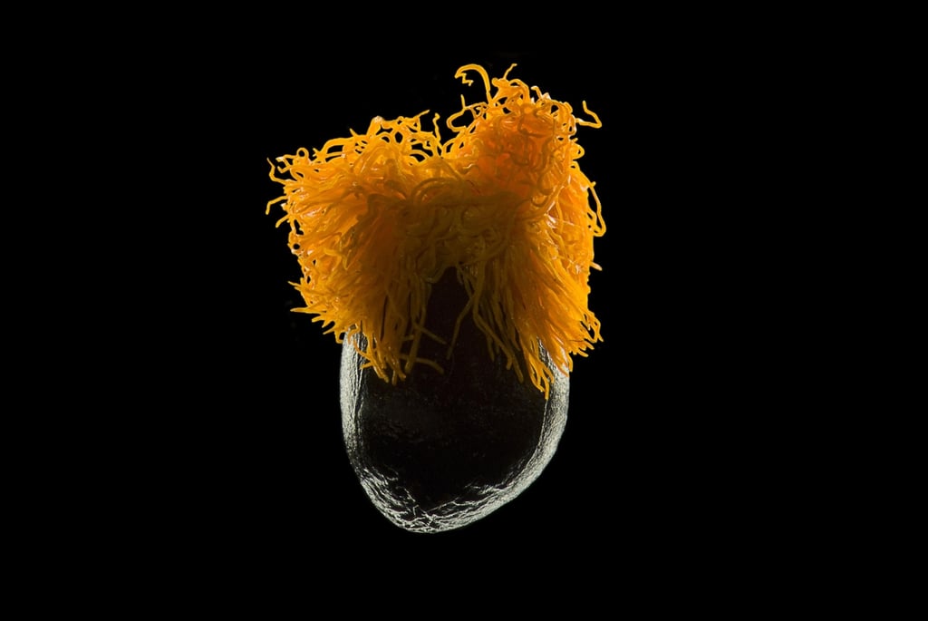
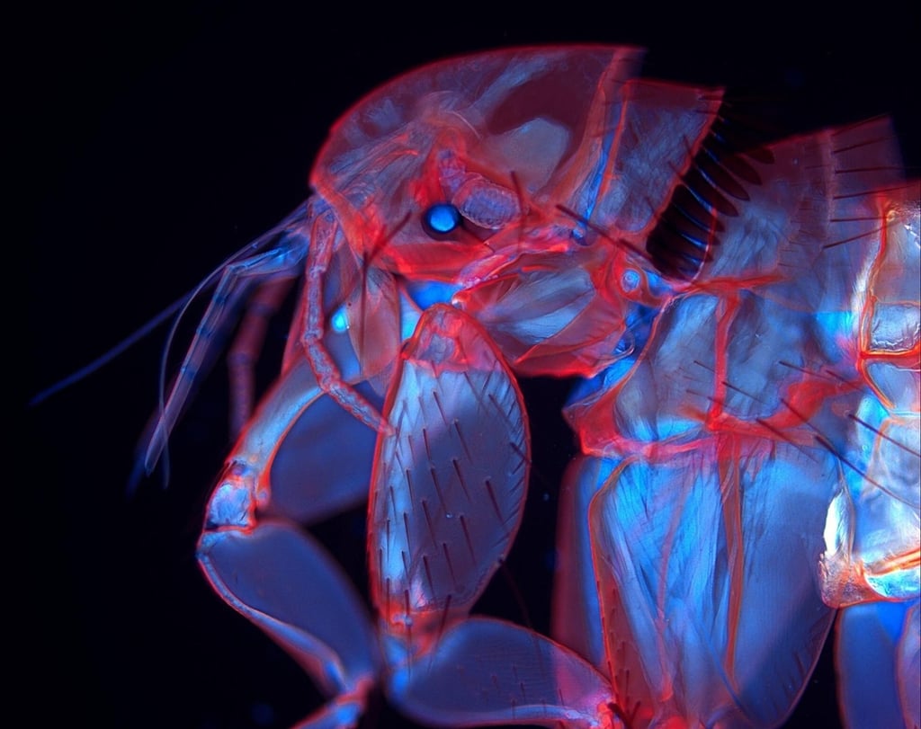
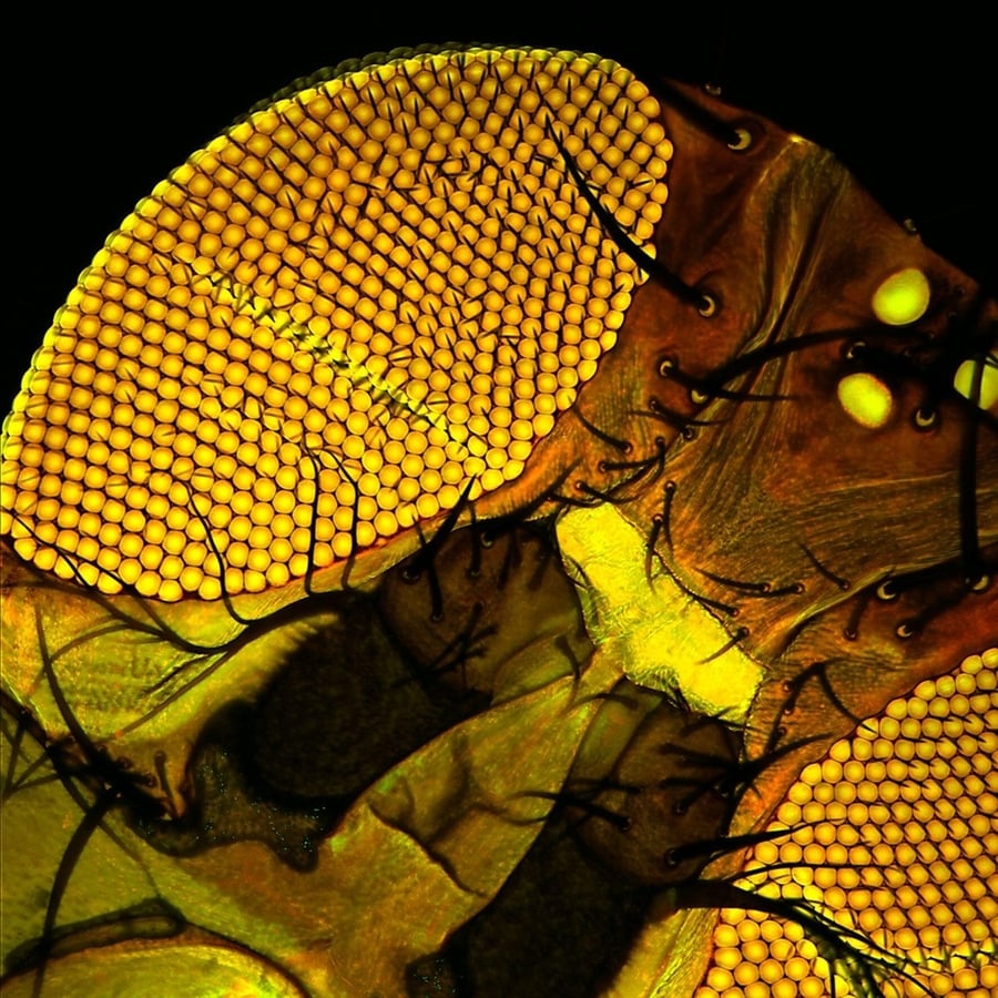
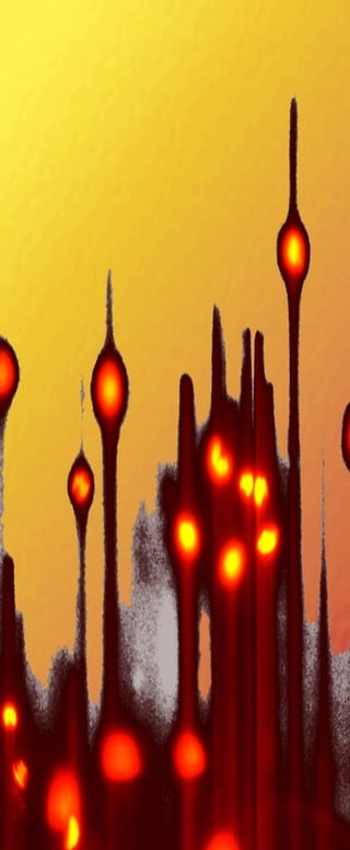
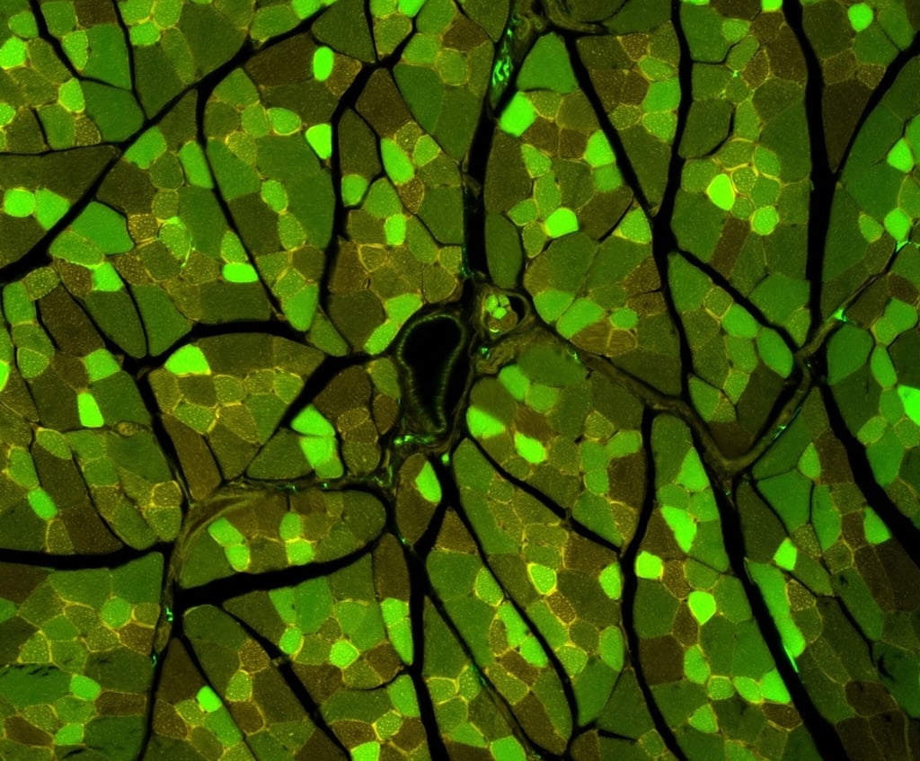
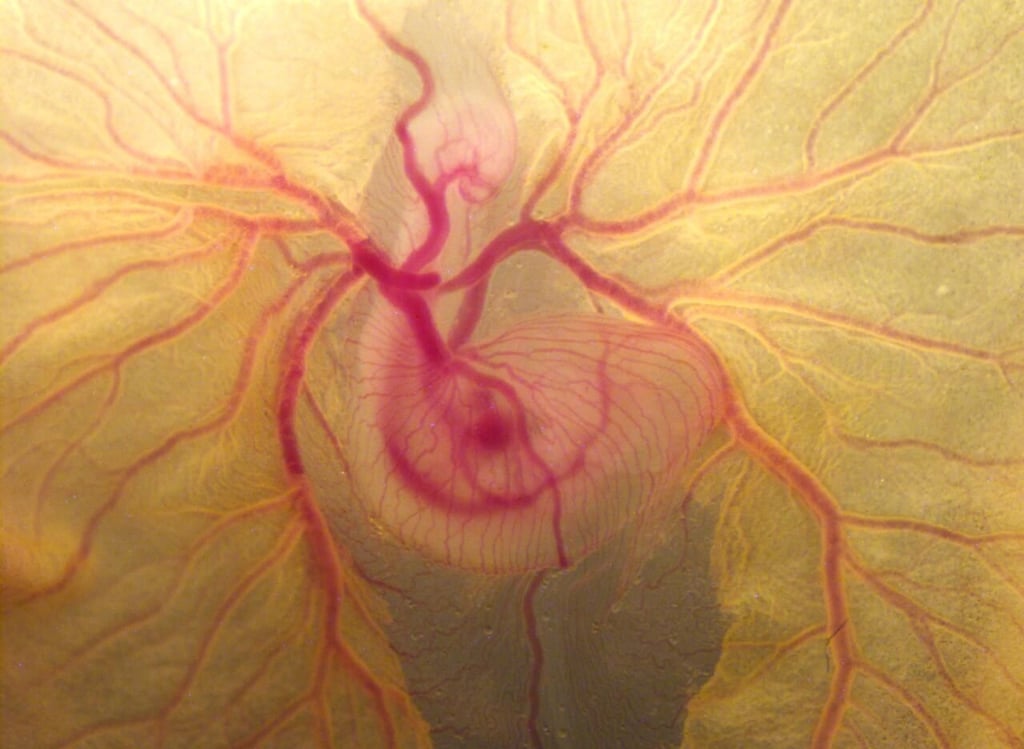
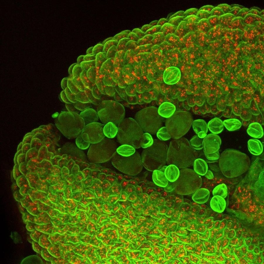
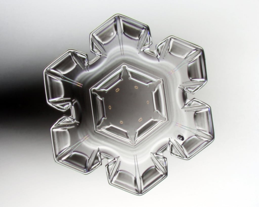
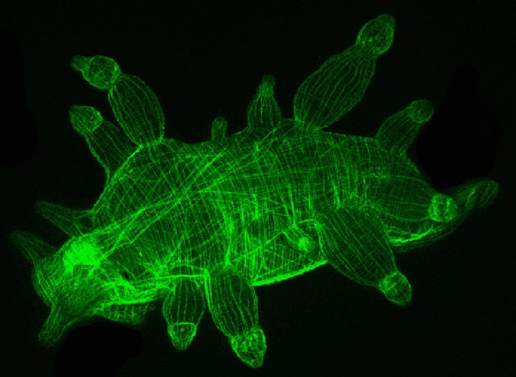
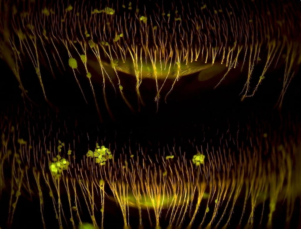
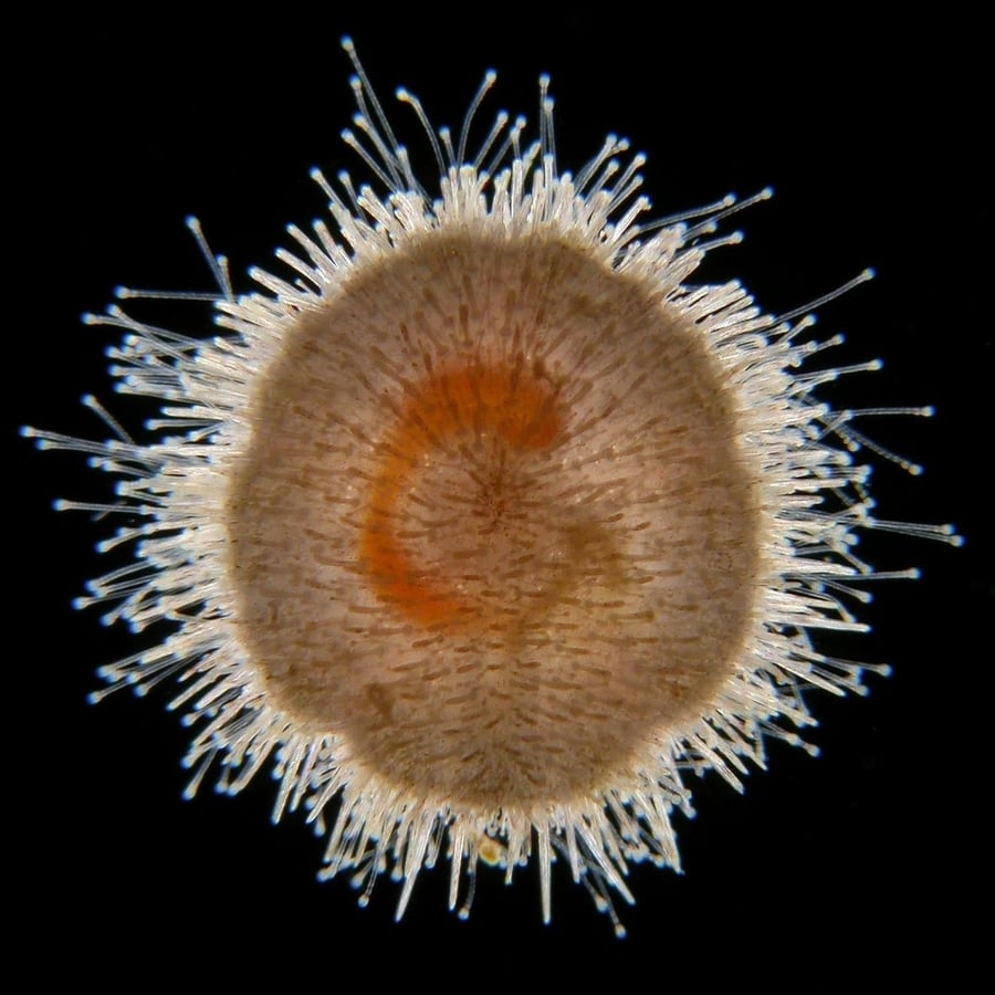
Oliver Braubach – Department of Physiology & Biophysics, Dalhousie University in Halifax, Nova Scotia, Canada
Courtesy of Nikon Small World
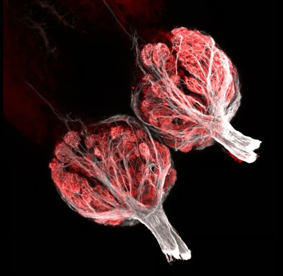
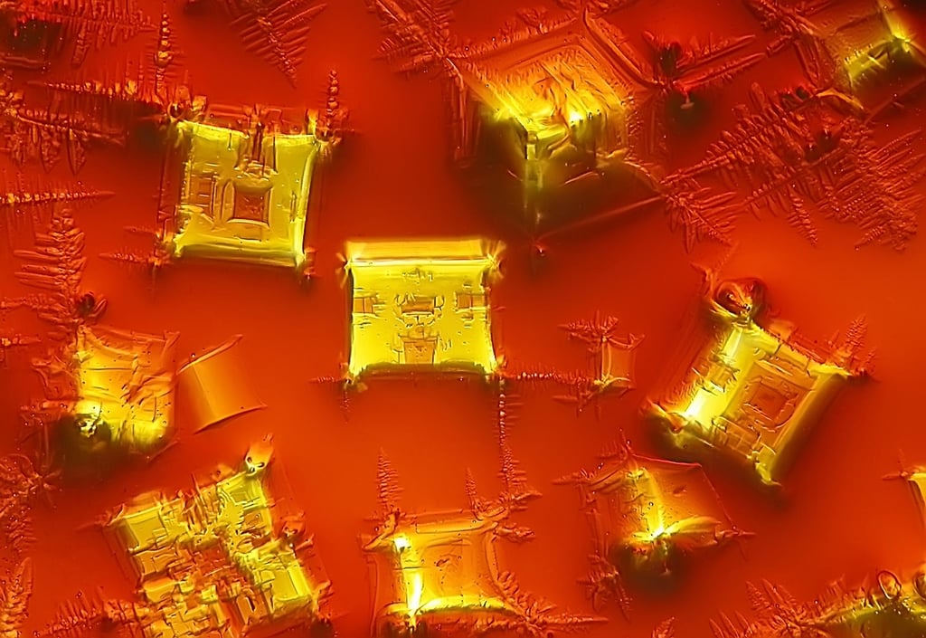
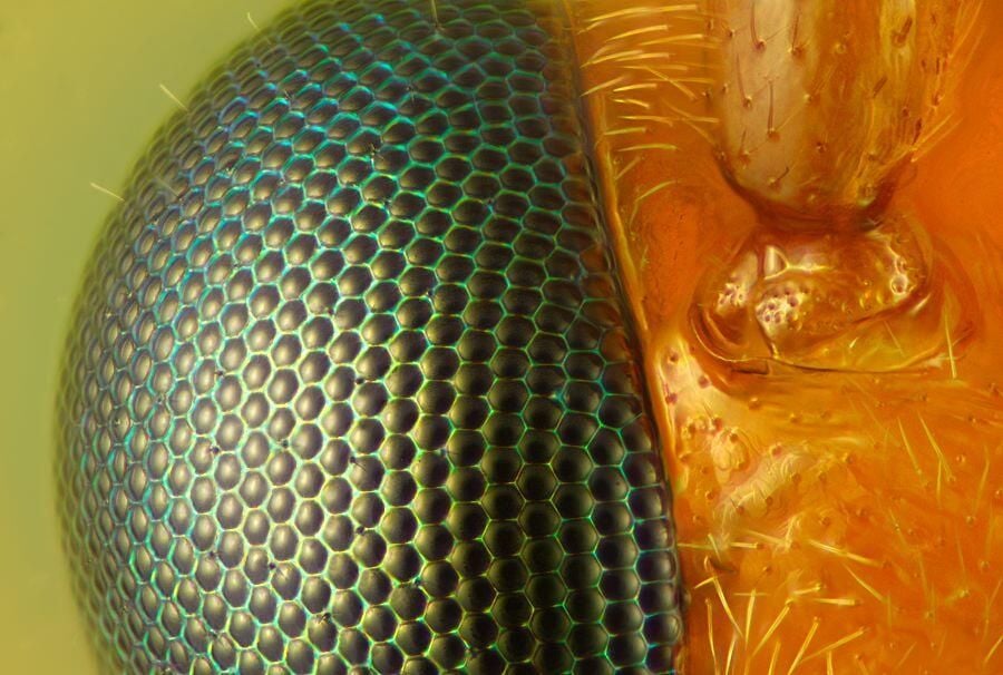
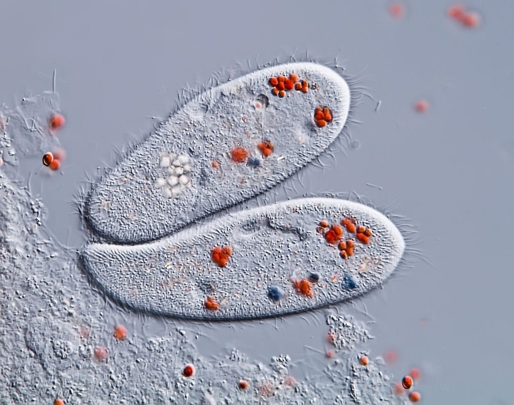
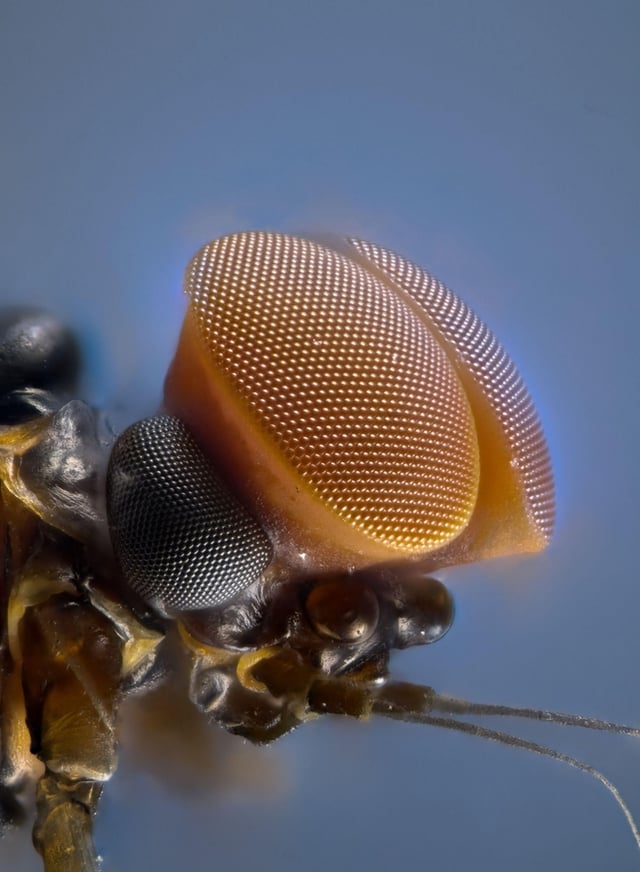
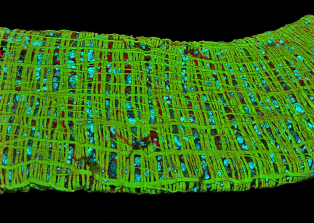
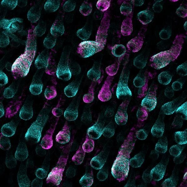
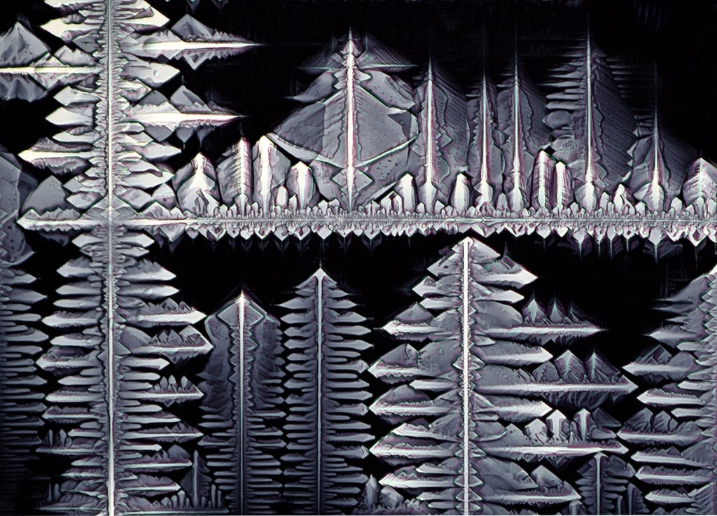
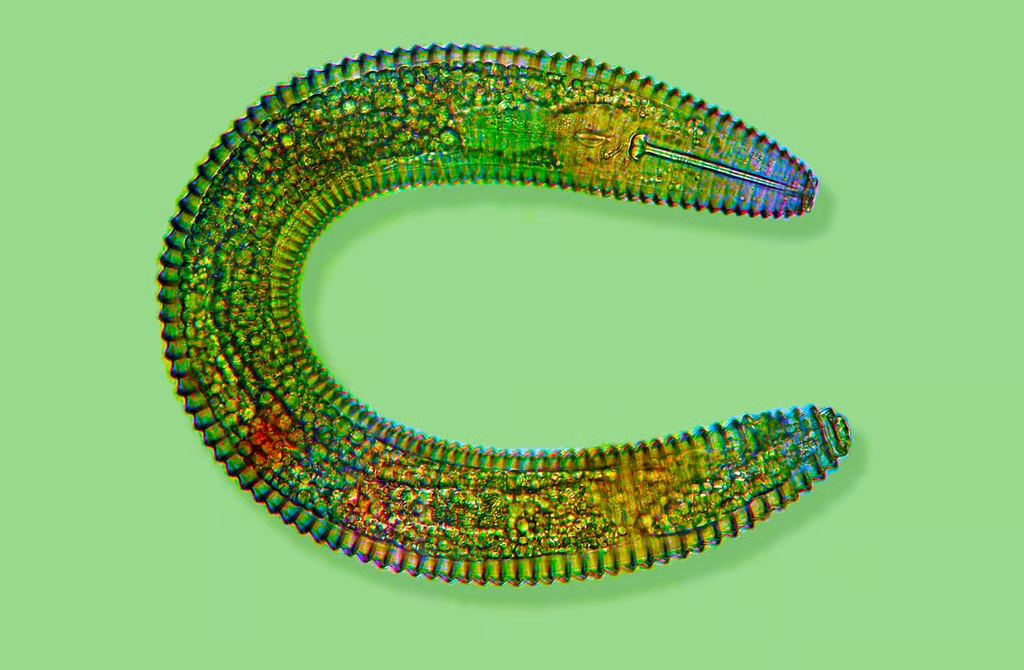
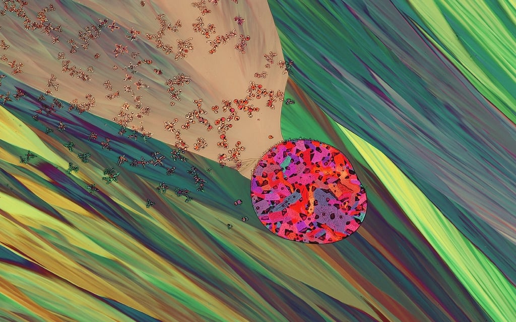
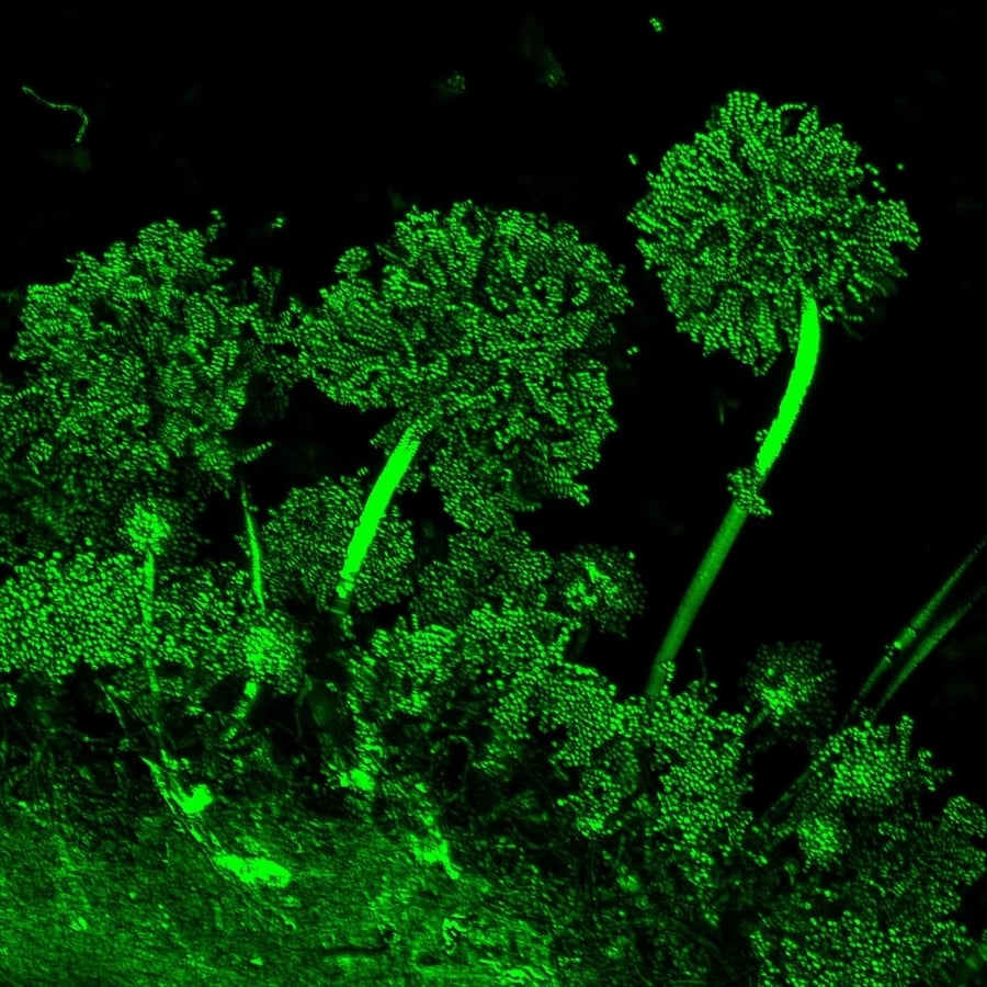
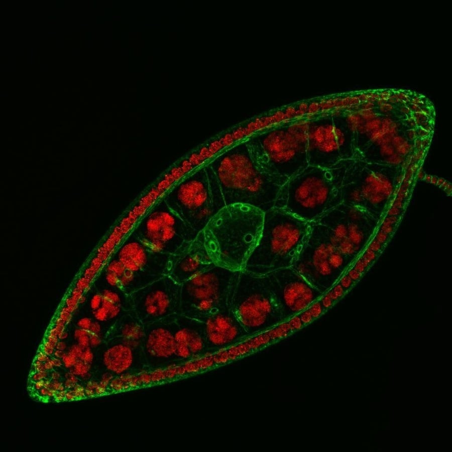
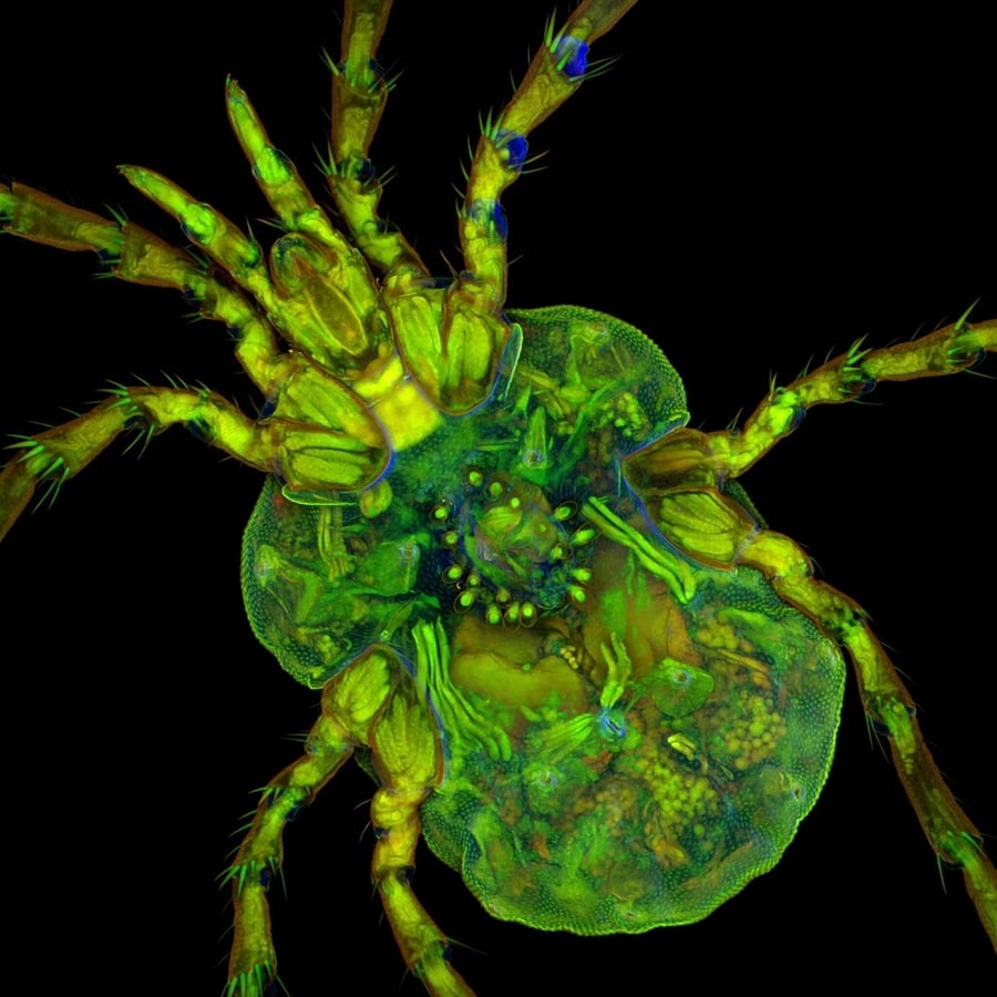
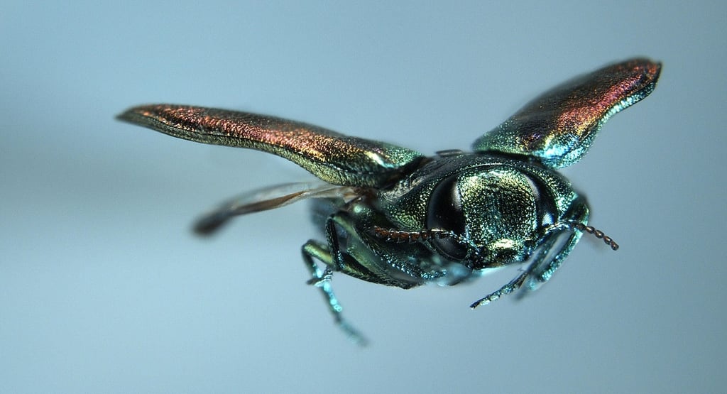
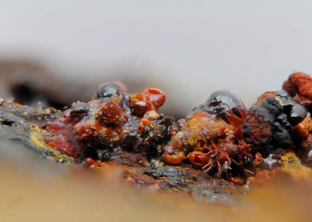
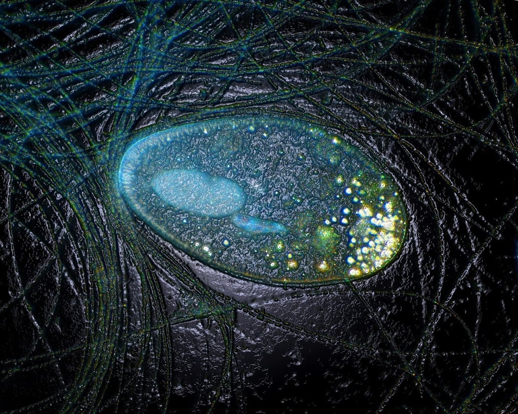
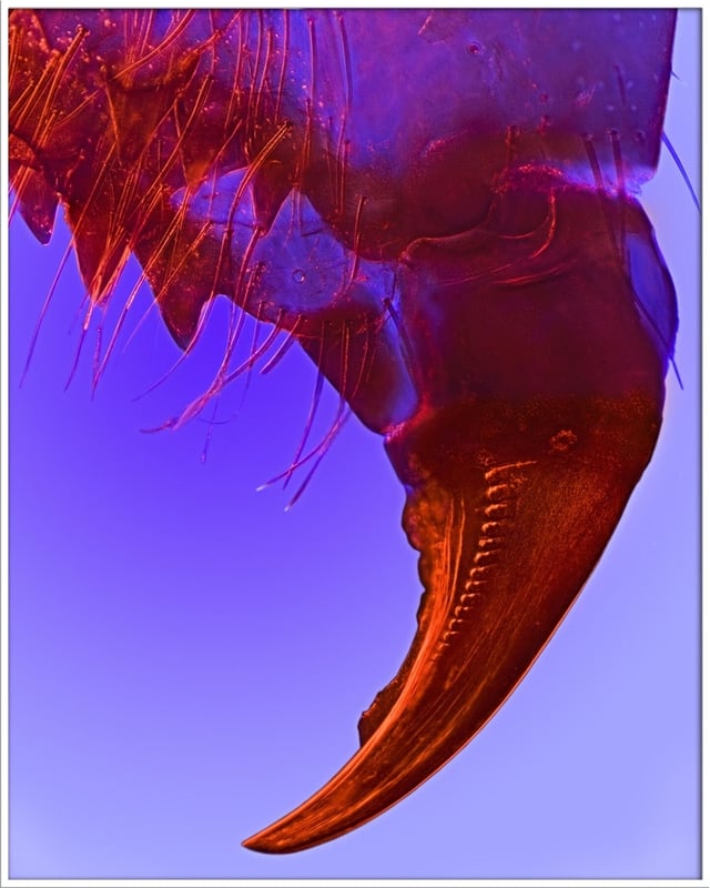
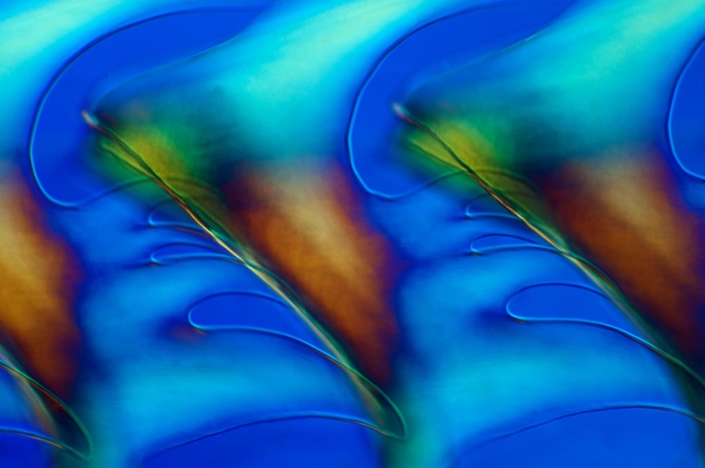
Riccardo Taiariol – La Spezia, SP, Italy
Courtesy of Nikon Small World
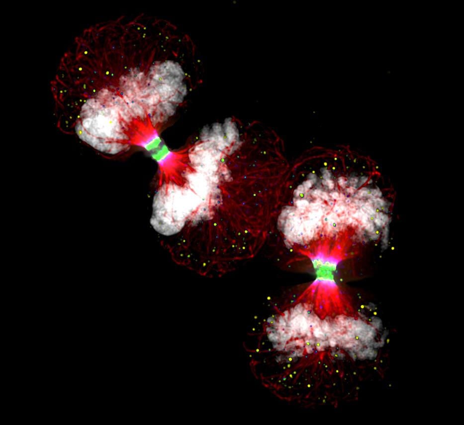
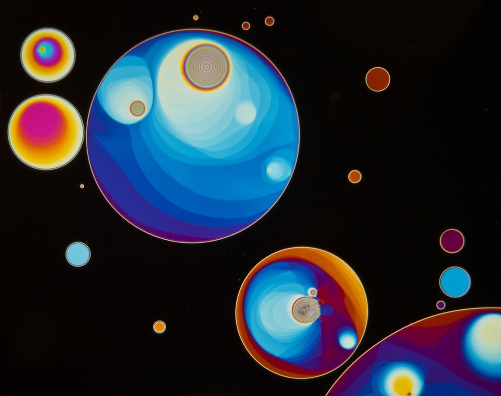
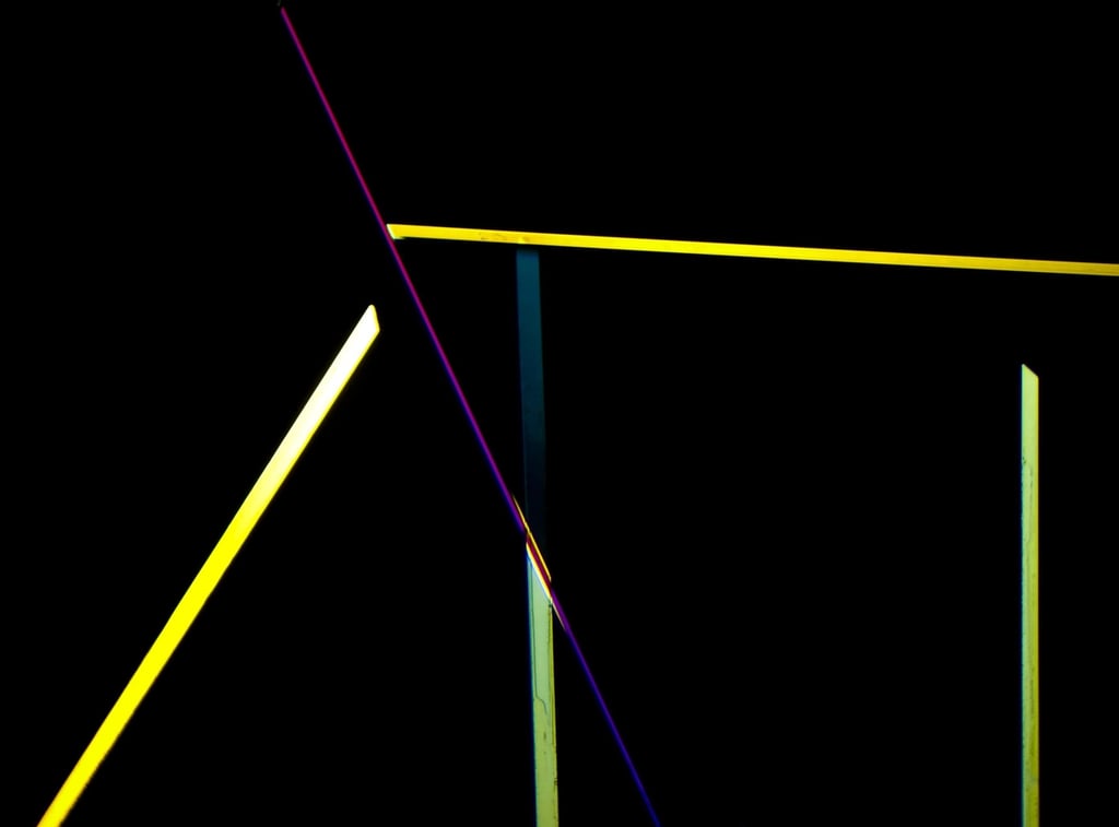
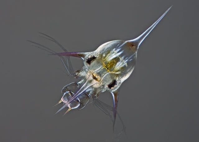
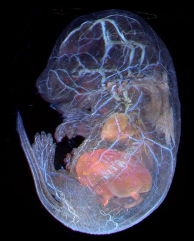
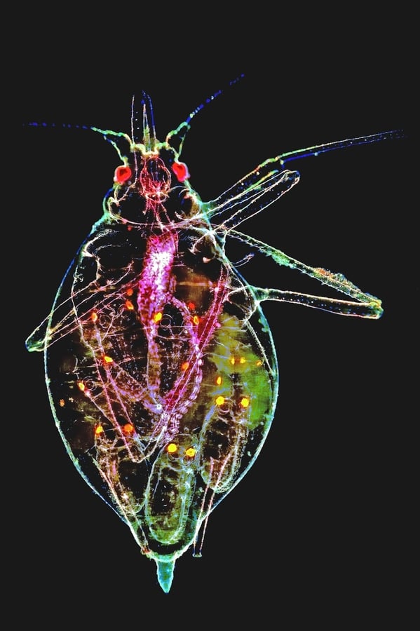
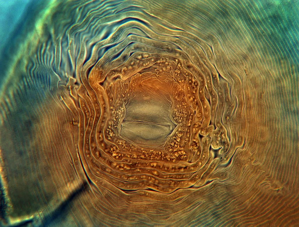
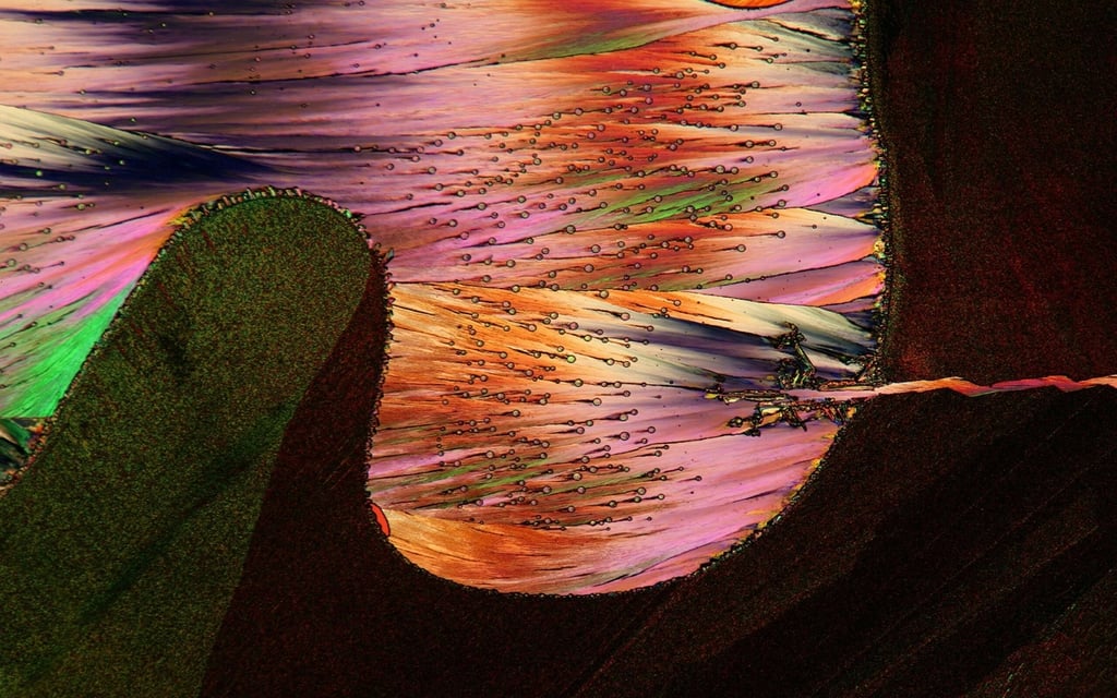
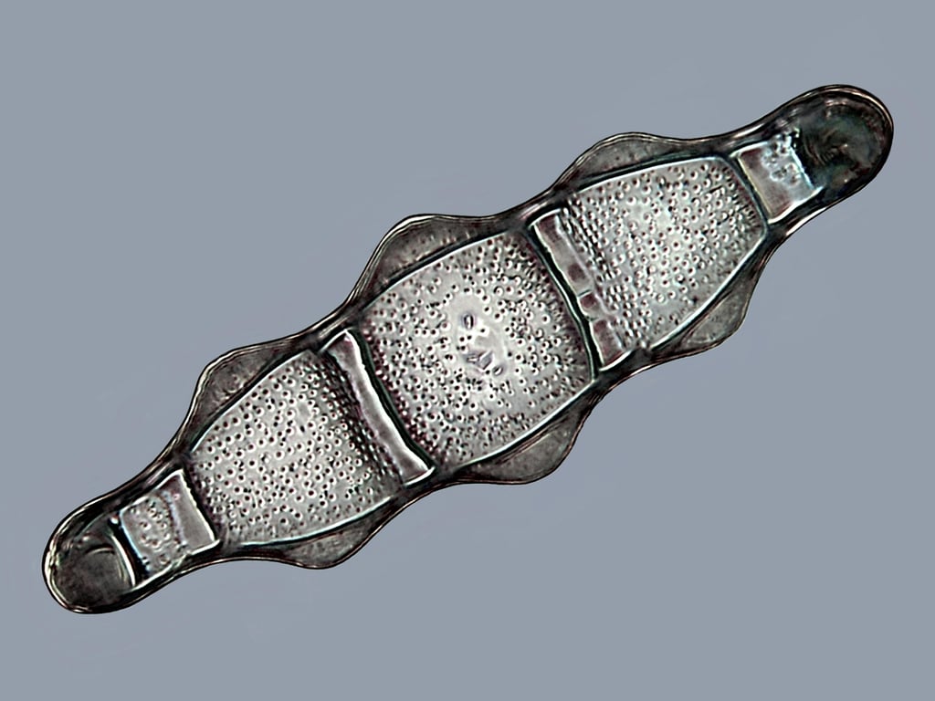
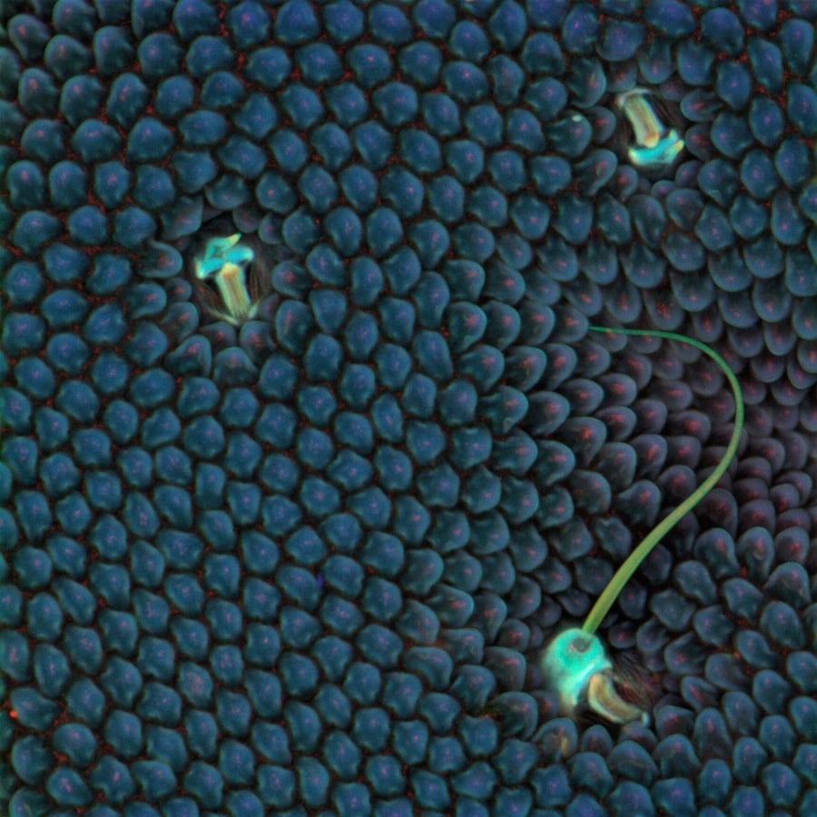
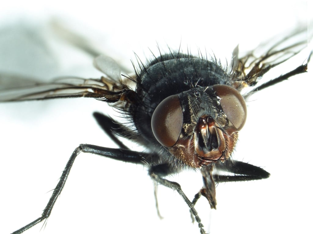
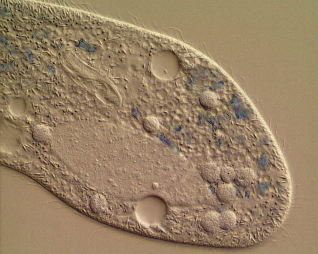
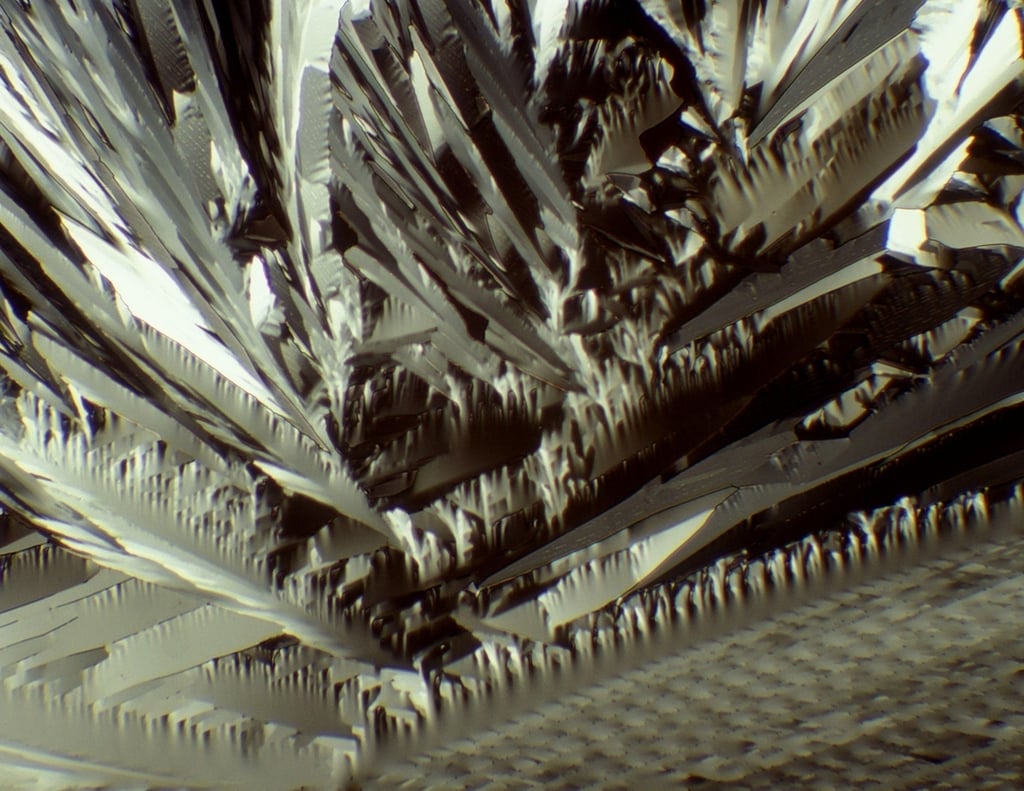
Viktor Sykora – Institute of Pathophysiology, First Medical Faculty, Charles University in Prague, Czech Republic
Courtesy of Nikon Small World
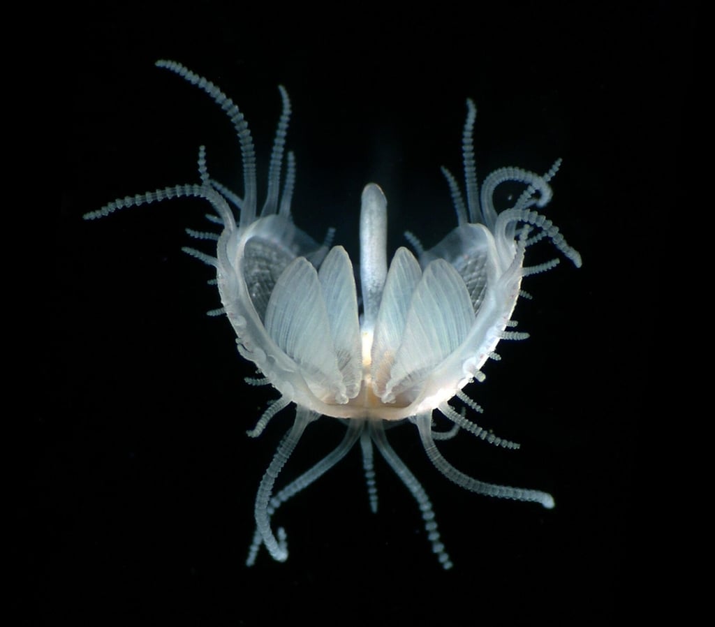
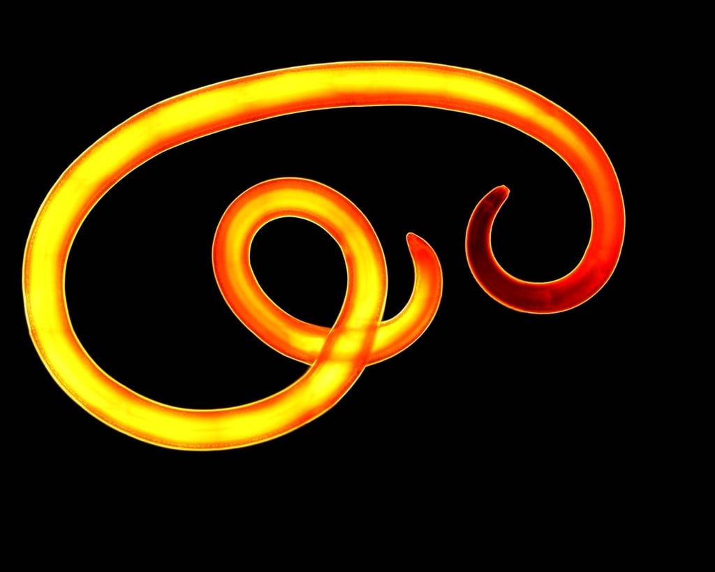
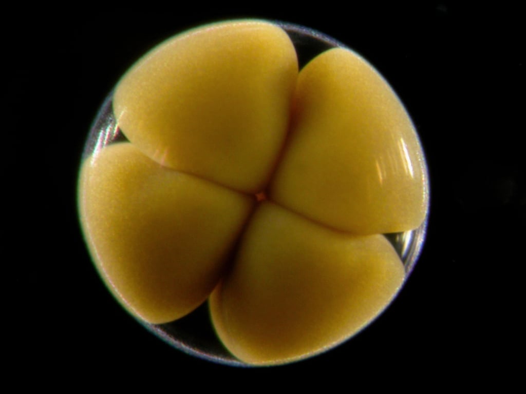
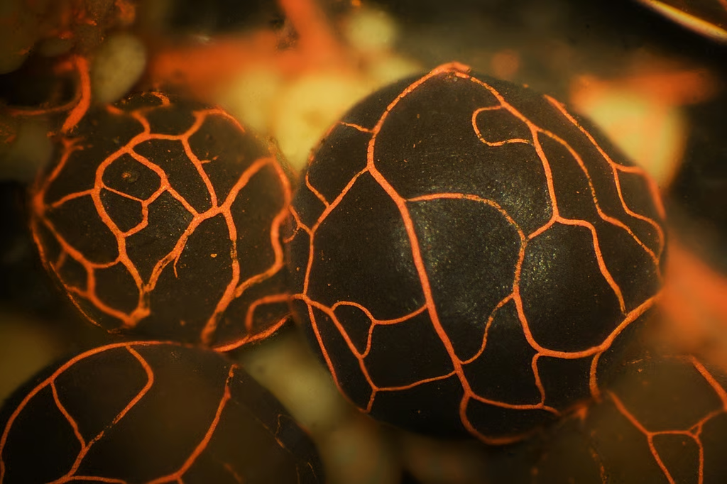
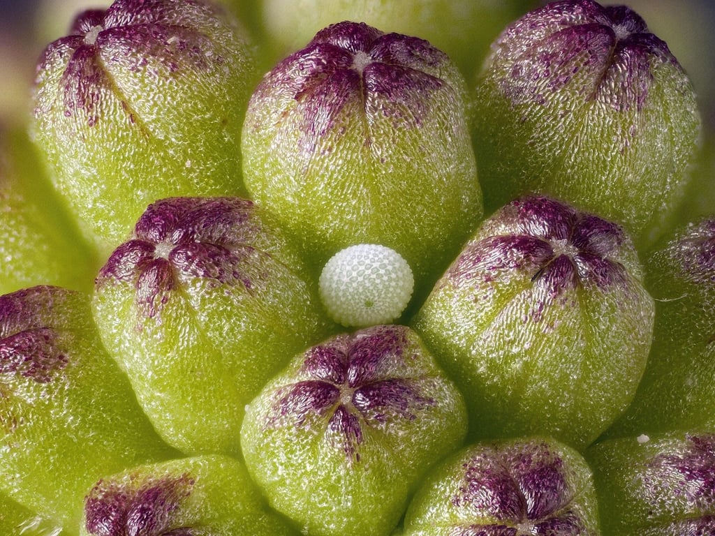
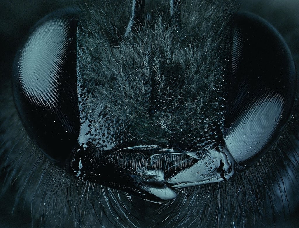
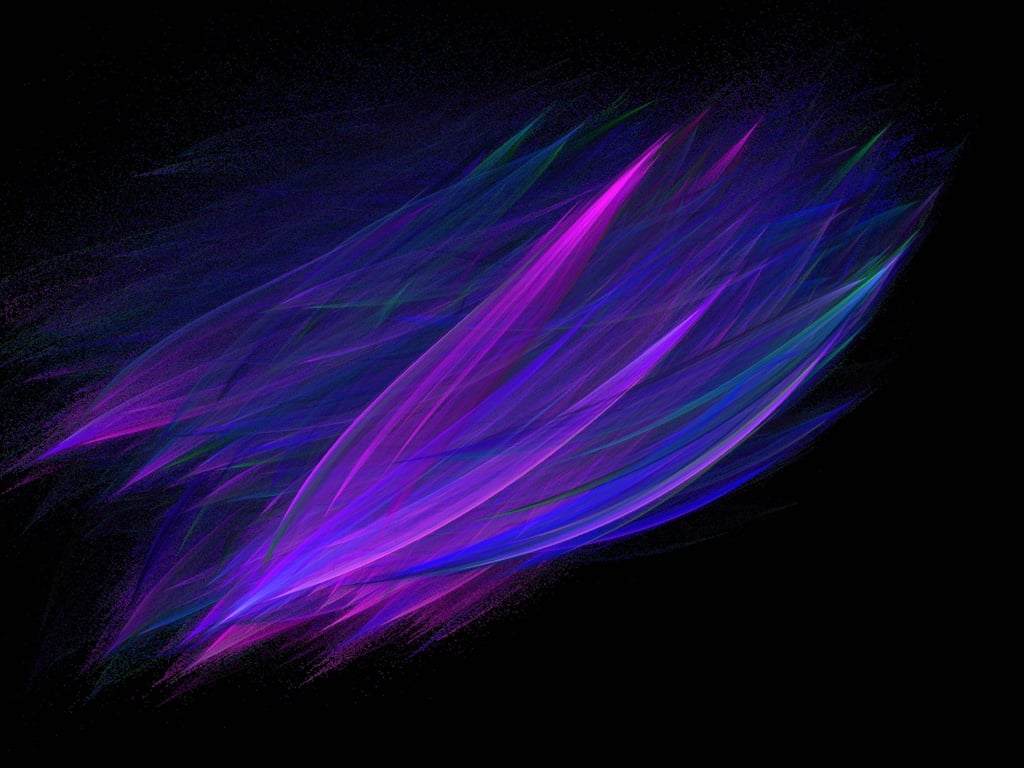
Dr. John Huisman – Murdoch University, School of Biological Sciences and Biotechnology in Murdoch, Western Australia, Australia
Courtesy of Nikon Small World
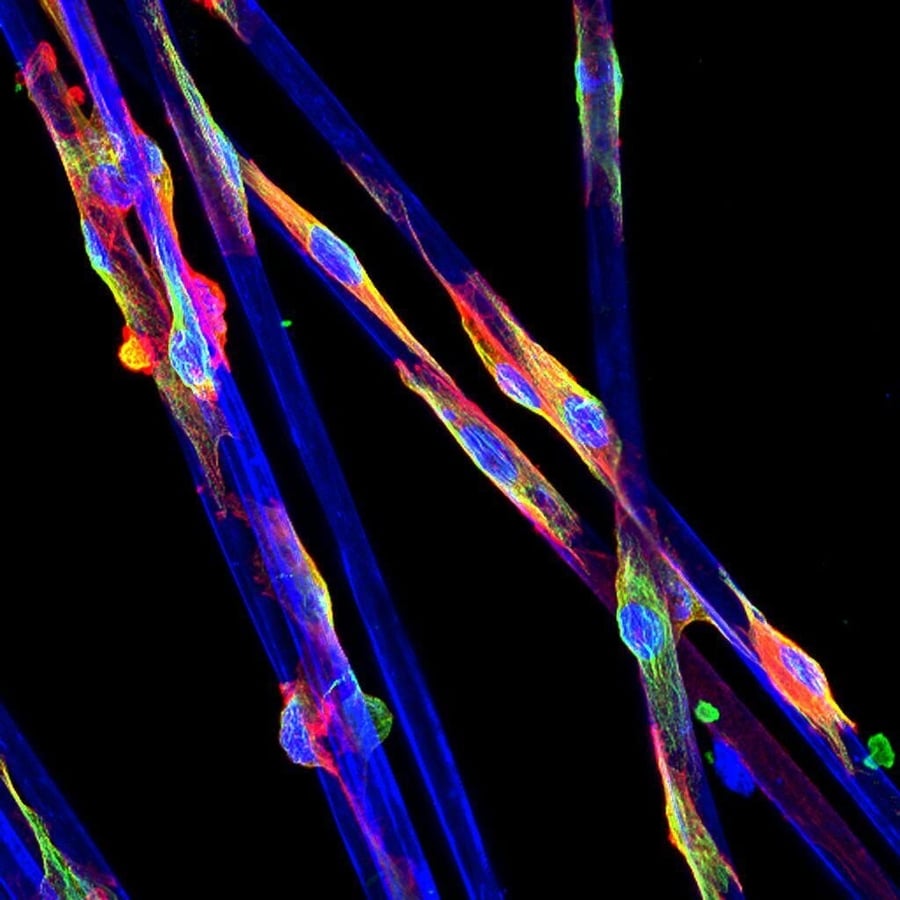
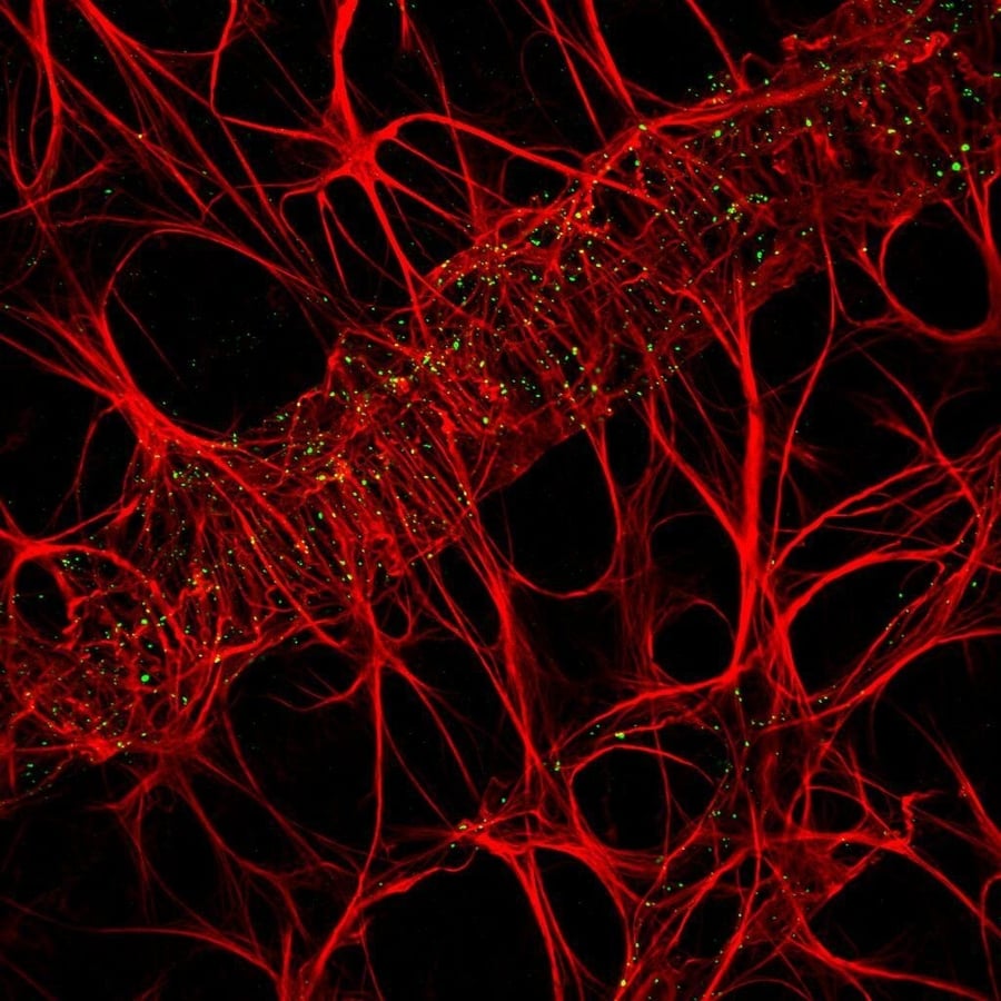
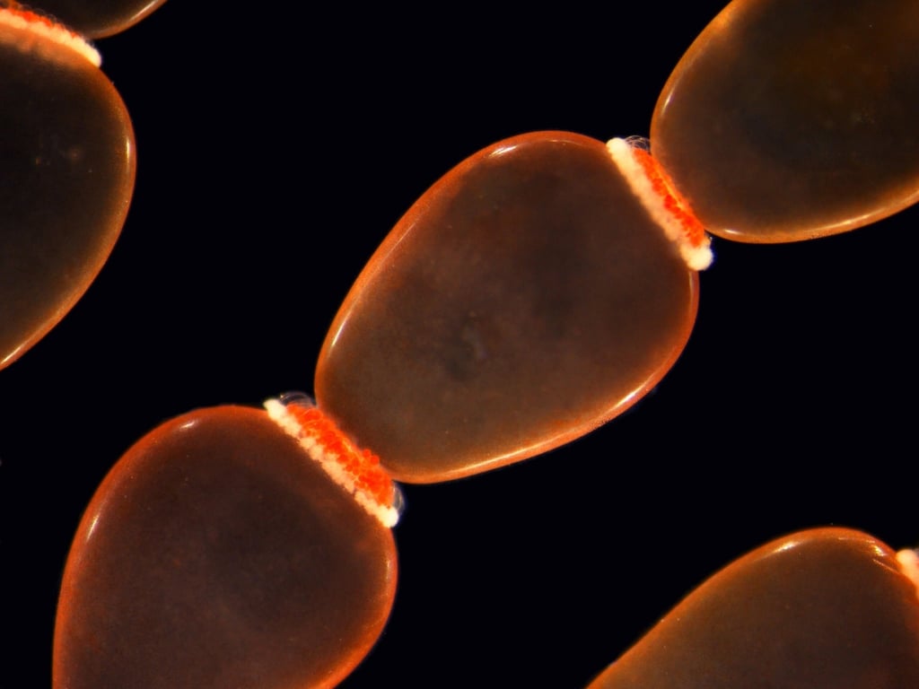
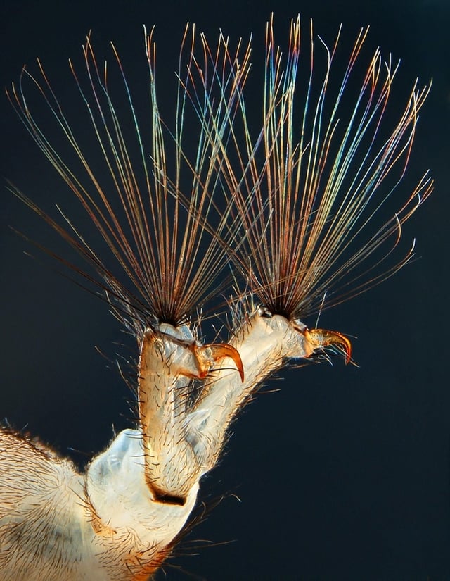
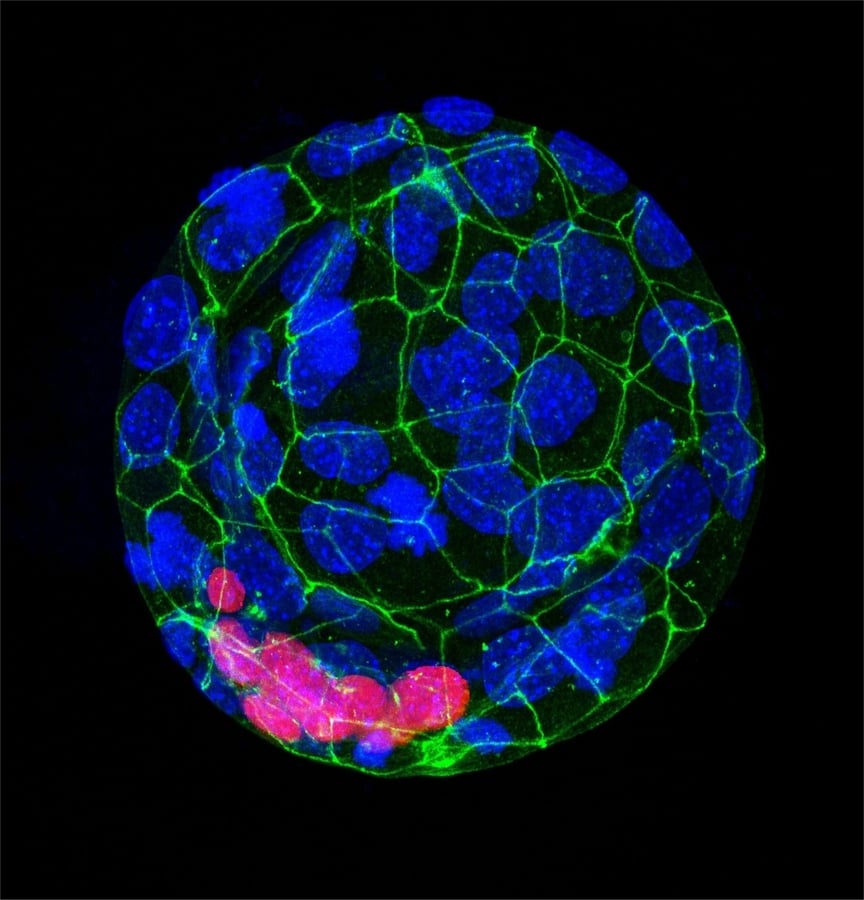
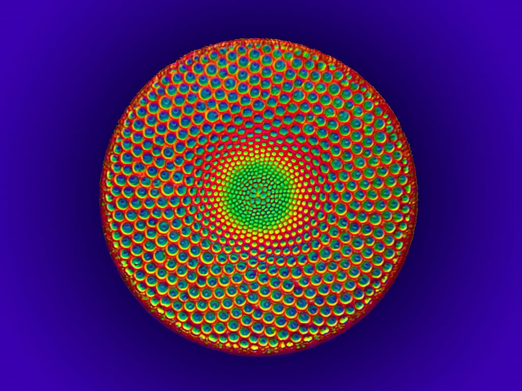
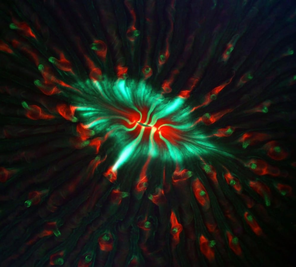
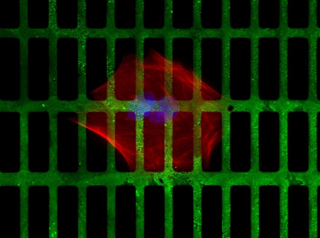
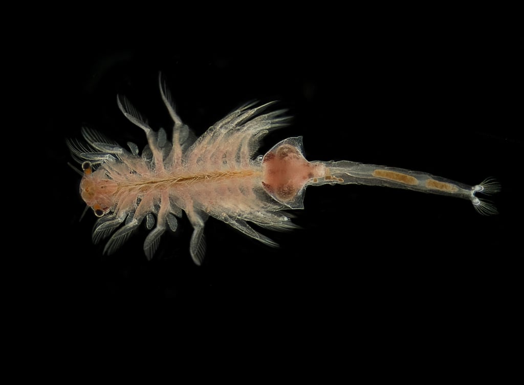
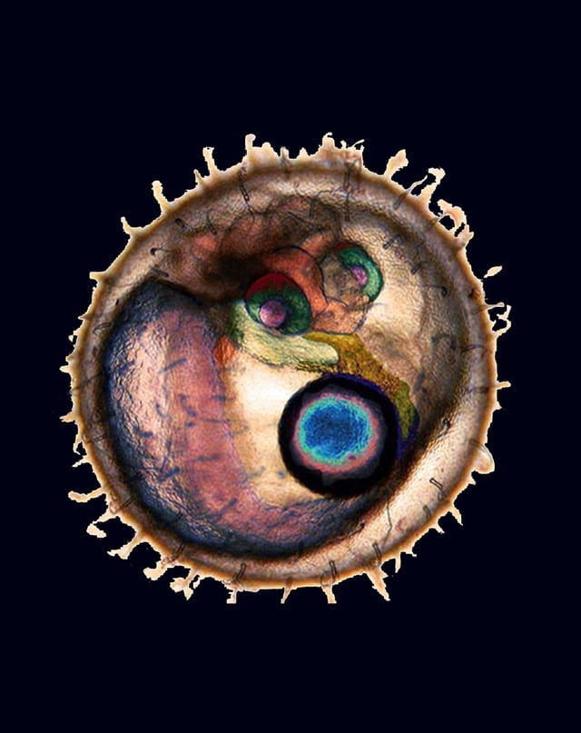
Yongli Shan – The University of Texas Southwestern Medical Center in Dallas, Texas
Courtesy of Nikon Small World
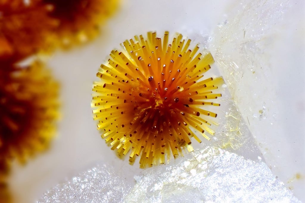
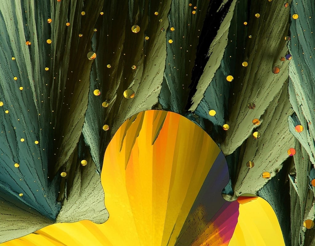
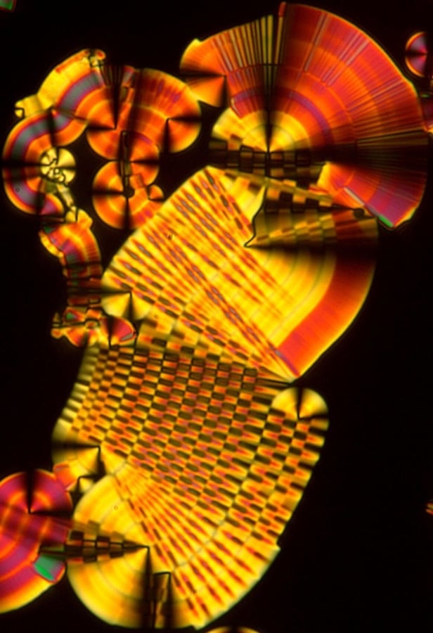
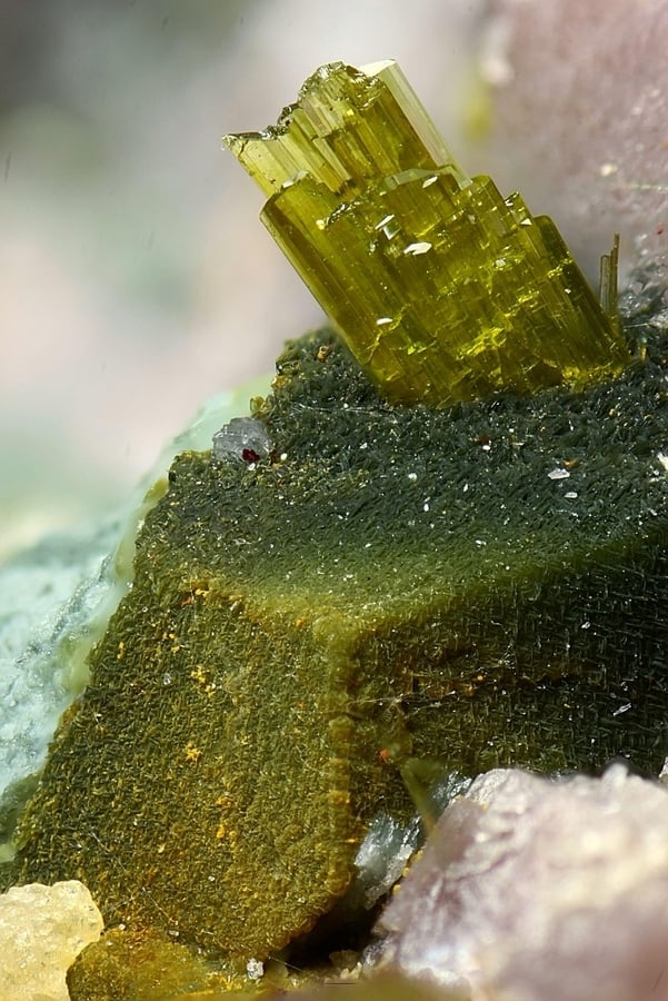
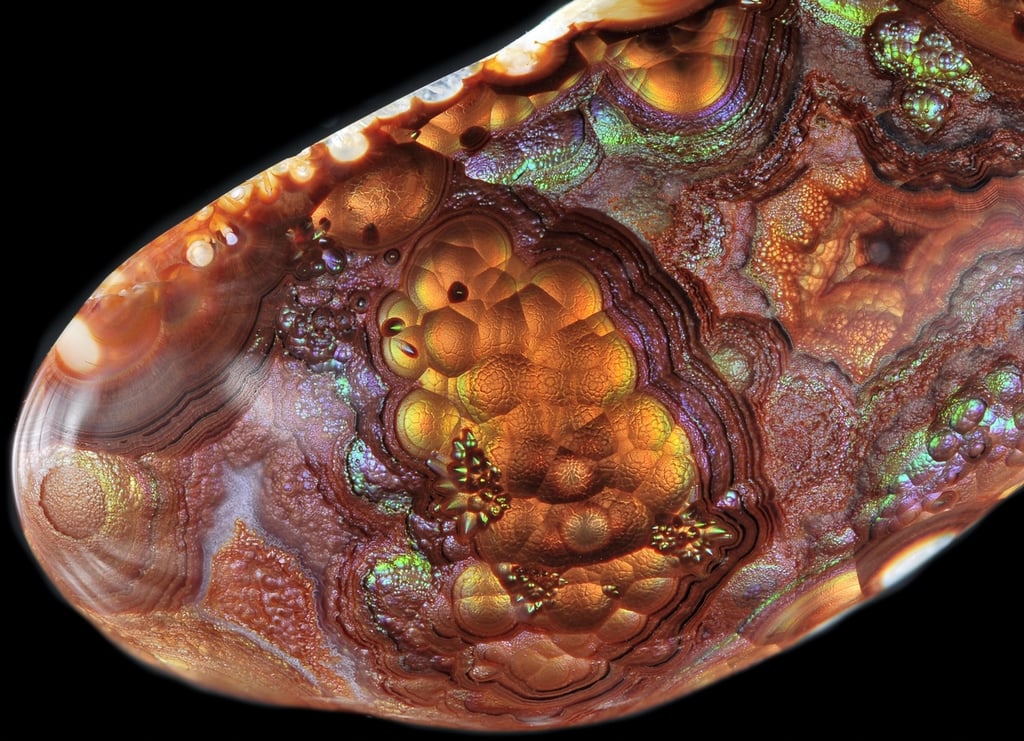
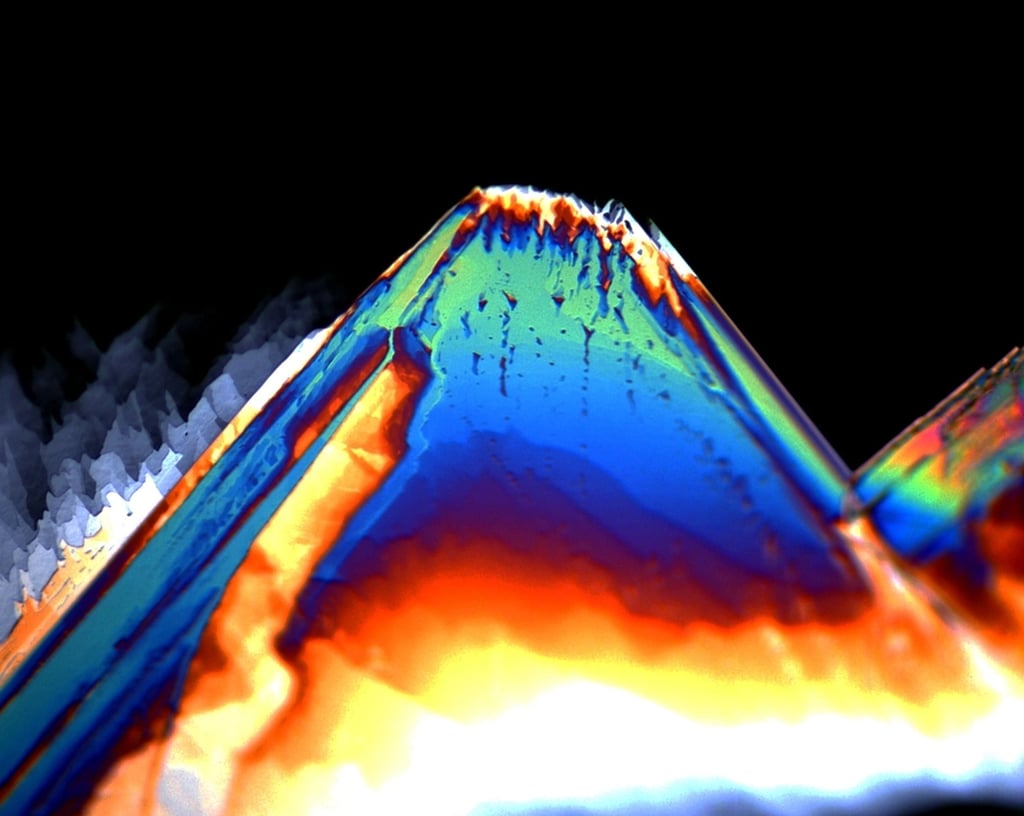
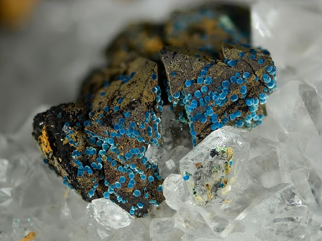
Honorio Cocera-La Parra – Geology Museum, University of Valencia in Benetusser, Valencia, Spain
Courtesy of Nikon Small World
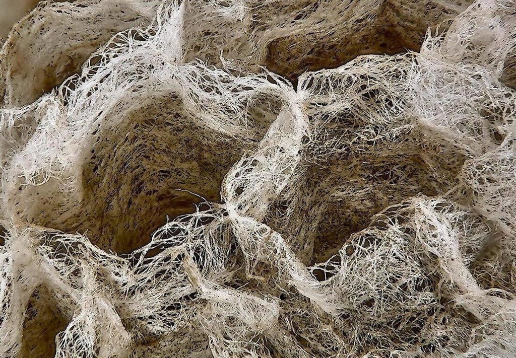
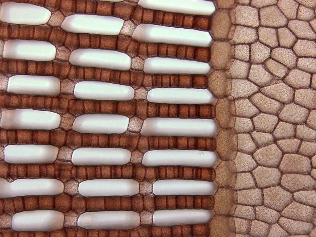
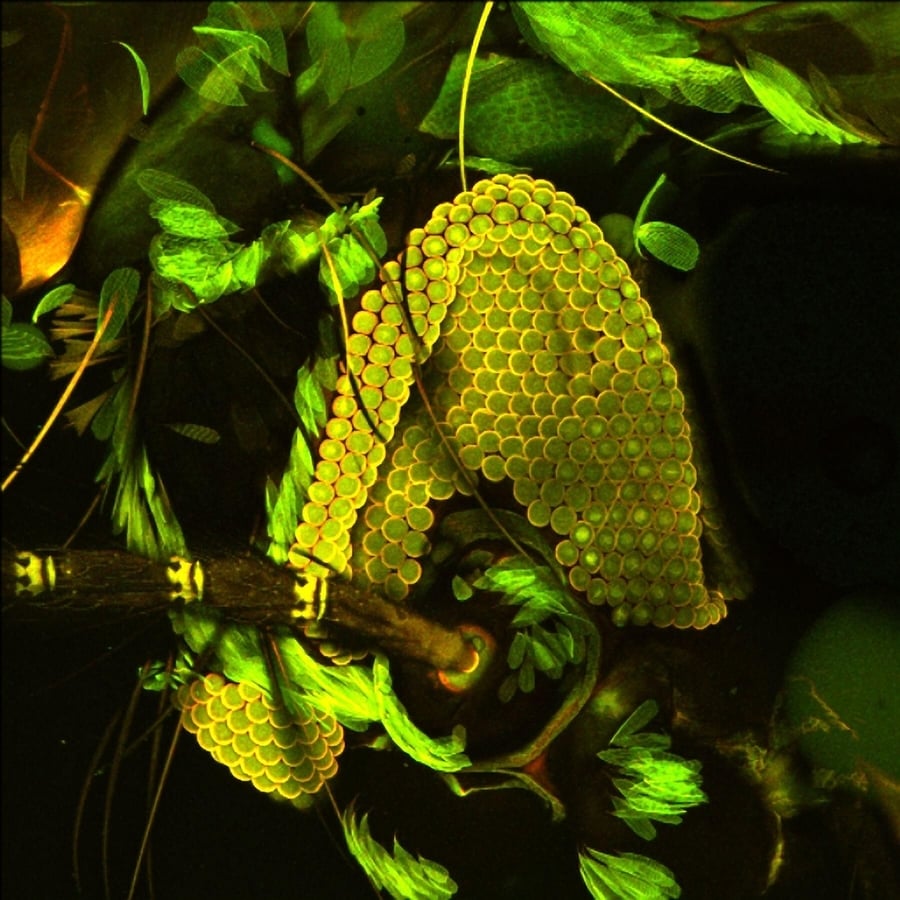
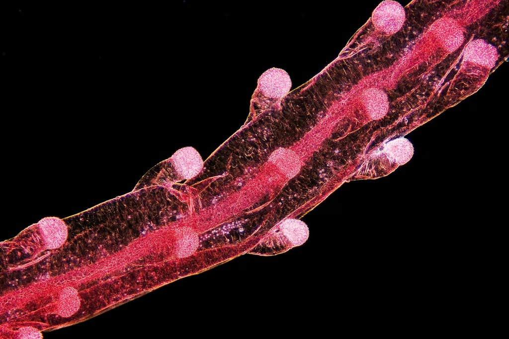
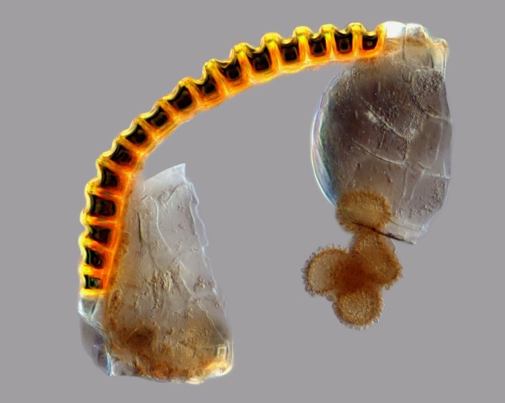
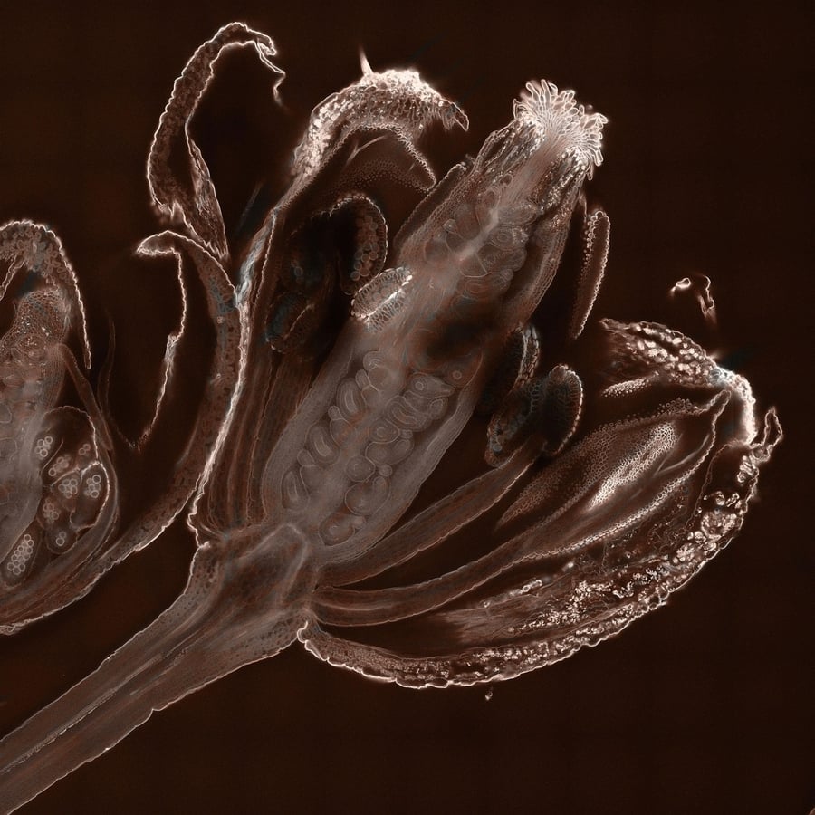
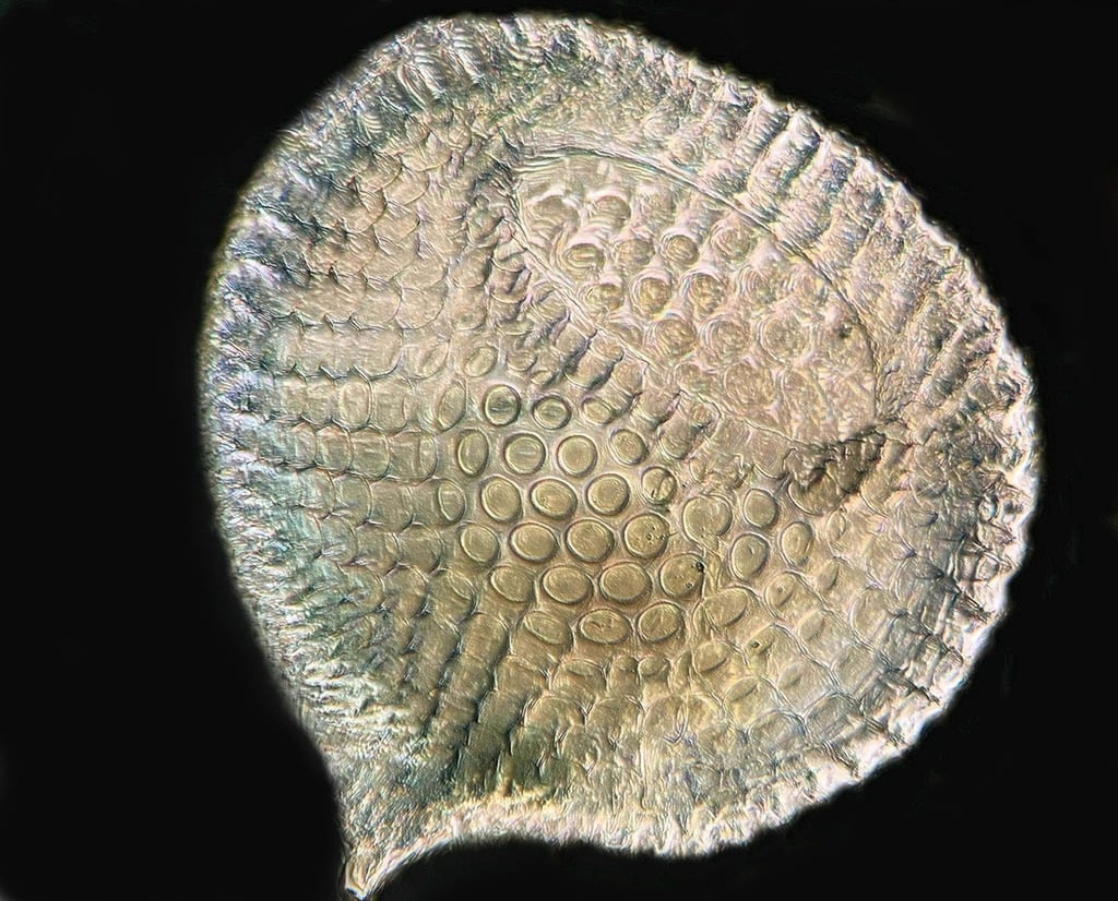
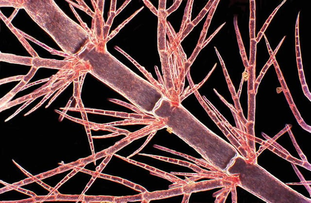
Dr. Duane Harland – AgResearch Ltd. in Lincoln, New Zealand
Courtesy of Nikon Small World
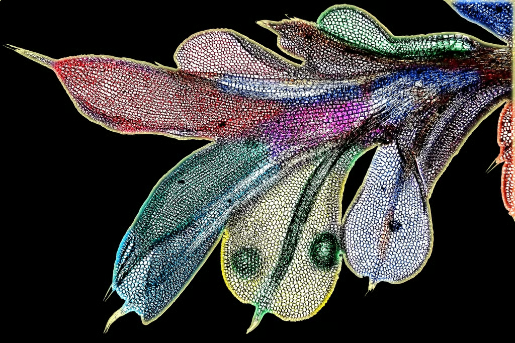
Yanping Wang – Beijing Language and Culture University in Beijing, China
Courtesy of Nikon Small World
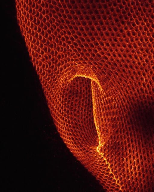
Dr. Paul D. Andrews – University of Dundee in Dundee, Scotland, UK
Courtesy of Nikon Small World
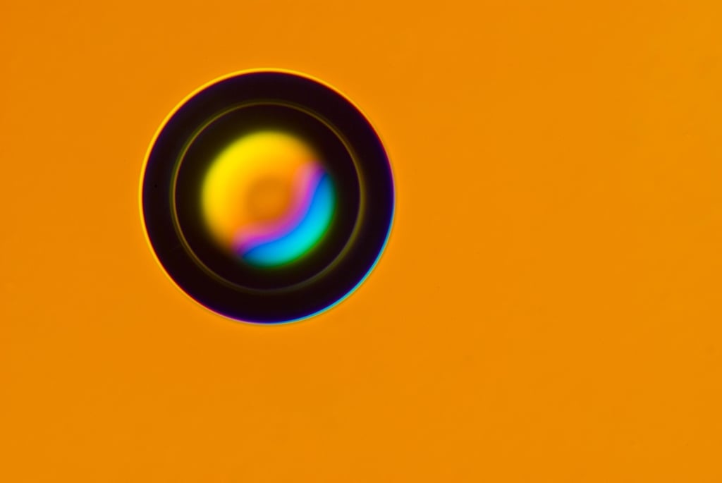
Dr. Gregory Rouse – Scripps Institution of Oceanography in La Jolla, California
Courtesy of Nikon Small World
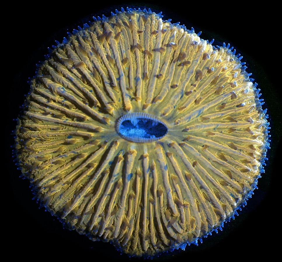
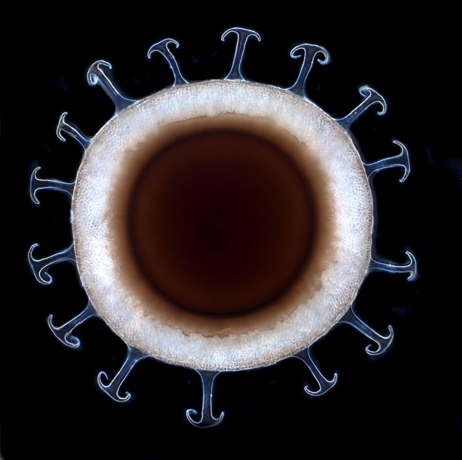
James Nicholson – NOAA NOS NCCOS Coral Culture and Collaborative Research Facility in Charleston, South Carolina
Courtesy of Nikon Small World
Dr. Stephen Lowry – University of Ulster in Portstewart, Co. Londonderry, UK
Courtesy of Nikon Small World
Dr. Ralf Wagner – D\u00fcsseldorf, Germany
Courtesy of Nikon Small World
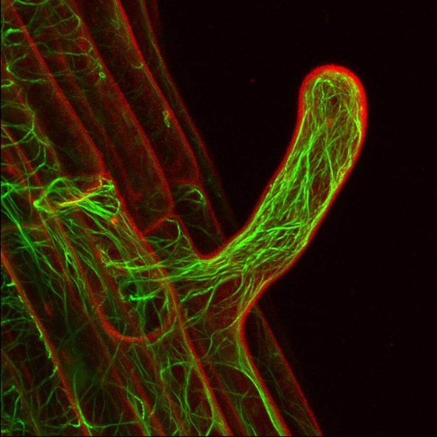
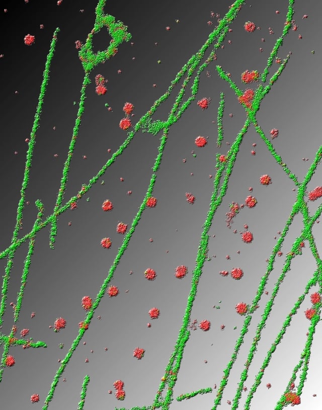
Dr. Robert Markus – Institute of Genetics, Biological Research Center of the Hungarian Academy of Sciences in Szeged, Hungary
Courtesy of Nikon Small World
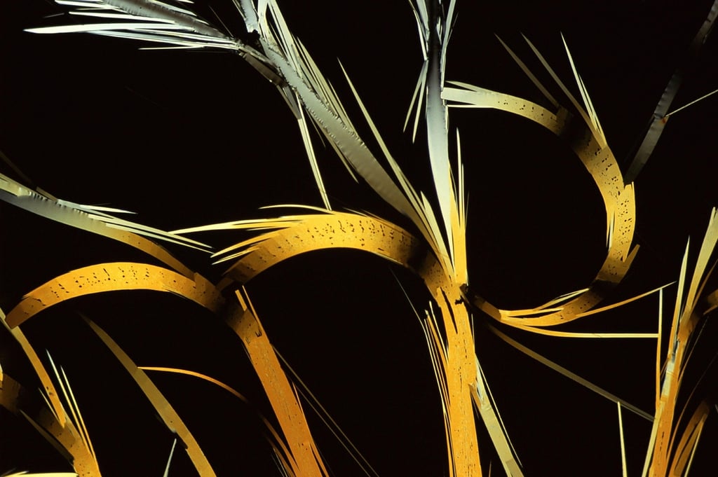
Charles Krebs – Charles Krebs Photography in Issaquah, Washington
Courtesy of Nikon Small World
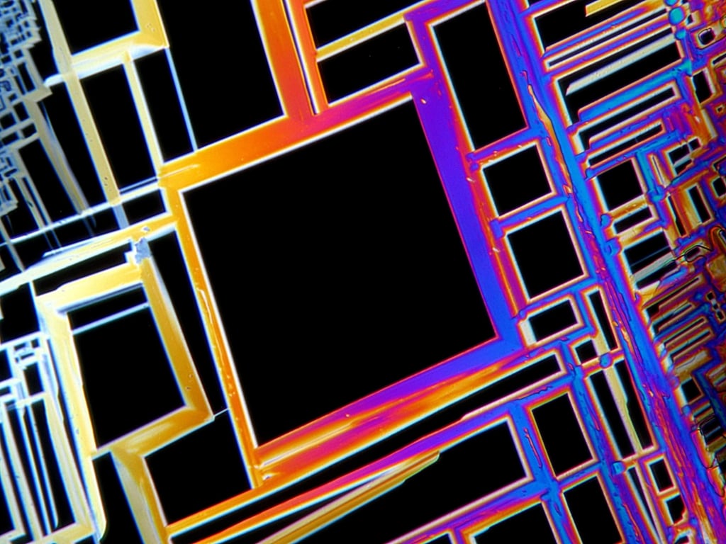
Gerd Guenther – D\u00fcsseldorf, NRW, Germany
Courtesy of Nikon Small World
Cameron Johnson – The University of Auckland in Auckland, New Zealand
Courtesy of Nikon Small World
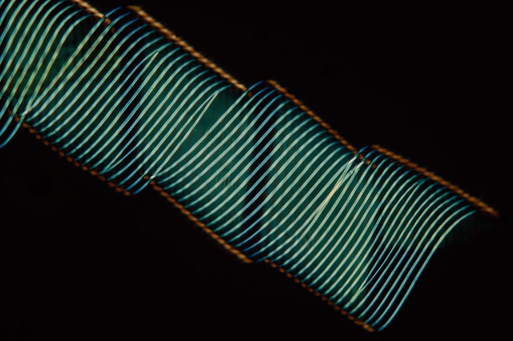
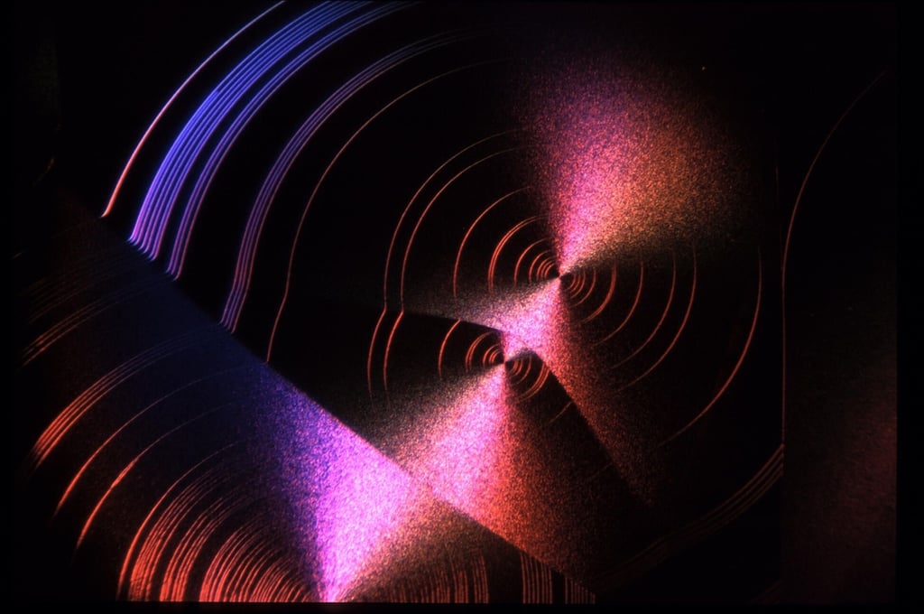
Dr. John Hart – Hart3D Films and Dept. Atmospheric and Oceanic Sci. Univ. in Boulder, Colorado
Courtesy of Nikon Small World
Dr. Marie Andersson – UCMR/Dep. Molecular Biology, Ume\u00e5 Universitet in Ume\u00e5, Sweden
Courtesy of Nikon Small World
Dr. Robert Berdan – Science & Art Multimedia in Calgary, Alberta, Canada
Courtesy of Nikon Small World
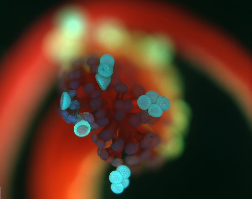
Rajdeep Deb – Assam University, Department of Chemistry in Assam, India
Courtesy of Nikon Small World
Dr. Edward Leighman Gafford – Ventura, California
Courtesy of Nikon Small World
Gerd Guenther – D\u00fcsseldorf, NRW, Germany
Courtesy of Nikon Small World
Darryl Hover – Salem, Oregon
Courtesy of Nikon Small World
J. Claire Hoving – Institute of Infectious Disease and Molecular Medicine, University Of Cape Town in Cape Town, Western Cape, South Africa
Courtesy of Nikon Small World
Dr. John Huisman – Murdoch University, School of Biological Sciences and Biotechnology in Murdoch, Western Australia, Australia
Courtesy of Nikon Small World
Laurie Knight – Tonbridge, Kent, UK
Courtesy of Nikon Small World
Charles Krebs – Charles Krebs Photography in Issaquah, Washington
Courtesy of Nikon Small World
Dr. Alvaro Migotto – Centro de Biologia Marinha, Universidade de S\u00e3o Paulo in S\u00e3o Paulo, SP, Brazil
Courtesy of Nikon Small World
Fabrice Parais – DREAL de Basse-Normandie in Caen, France
Courtesy of Nikon Small World
Dr. Mar\u00eda Prado-Figueroa – INIBIBB (CONICET – Universidad Nacional del Sur) in Bah\u00eda Blanca, Buenos Aires, Argentina
Courtesy of Nikon Small World
Antonio G. Valdecasas, Jose M. Becerra – Museo Nacional de Ciencias Naturales, CSIC in Madrid, Spain
Courtesy of Nikon Small World
Yanping Wang – Beijing Language and Culture University in Beijing, China
Courtesy of Nikon Small World
Rafael Pennese – Ecole Polytechnique F\u00e9d\u00e9rale de Lausanne in Lausanne, Vaud, Switzerland
Courtesy of Nikon Small World
Jose R. Almodovar – Biology Department, University of Puerto Rico Mayaguez Campus in Mayaguez, Puerto Rico
Courtesy of Nikon Small World
Jose R. Almodovar – Biology Department, University of Puerto Rico Mayaguez Campus in Mayaguez, Puerto Rico
Courtesy of Nikon Small World
Dr. Marie Andersson – UCMR/Dep. Molecular Biology, Ume\u00e5 Universitet in Ume\u00e5, Sweden
Courtesy of Nikon Small World
Dr. Paul Appleton, Ian Newton – University of Dundee in Dundee, Scotland, UK
Courtesy of Nikon Small World
Lars Bech – Naarden, The Netherlands
Courtesy of Nikon Small World
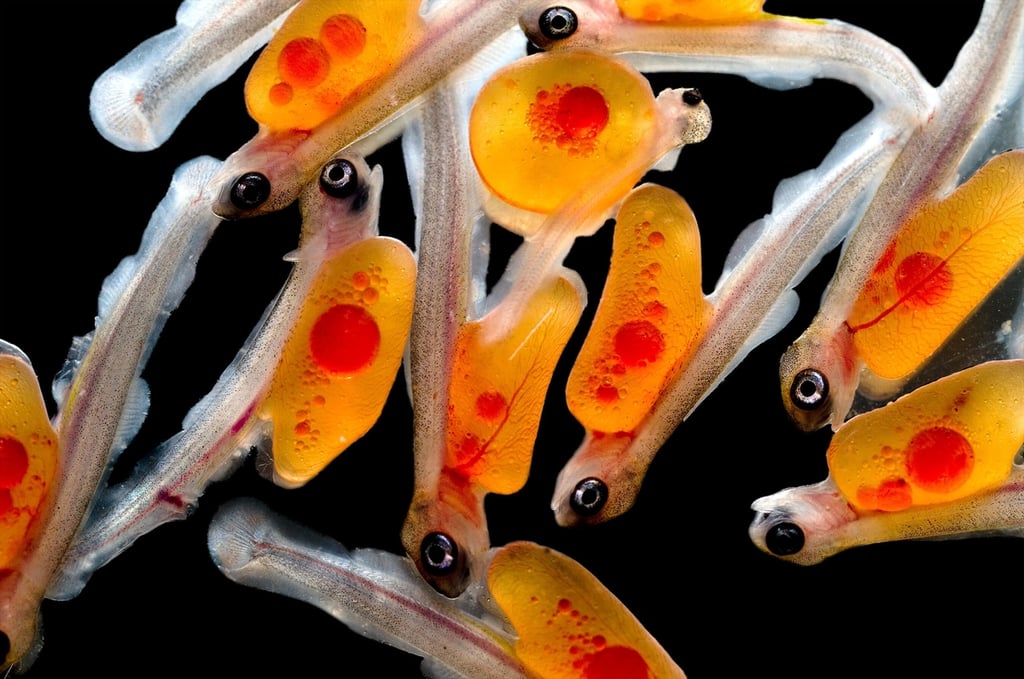
Tora Bardal – Department of Biology, NTNU Center of Fisheries and Aquaculture in Trondheim, Norway
Courtesy of Nikon Small World
Dr. Slobodan Beronja – The Rockefeller University in New York City, New York
Courtesy of Nikon Small World
Rachel Berry – Human Genetics Unit, MRC, Western General Hospital in Edinburgh, Scotland, UK
Courtesy of Nikon Small World
Dr. Elison Blancaflor – The Samuel Roberts Noble Foundation, Plant Biology in Ardmore, Oklahoma
Courtesy of Nikon Small World
Dr. Tomas Cabello – Universidad of Almer\u00eda in Roquetas de Mar, Spain
Courtesy of Nikon Small World
Jocelyn Cheng – Rochester Institute of Technology in Rochester, New York
Courtesy of Nikon Small World
Honorio Cocera-La Parra – Geology Museum, University of Valencia in Benetusser, Valencia, Spain
Courtesy of Nikon Small World
Thomas Deerinck – National Center for Microscopy and Imaging Research, University of California, San Diego – La Jolla, California
Courtesy of Nikon Small World
Thomas Deerinck – National Center for Microscopy and Imaging Research, University of California, San Diego – La Jolla, California
Courtesy of Nikon Small World
Dr. Laurence Dubreil – DPTM-UMR703-INRA-ONIRIS in Nantes, France
Courtesy of Nikon Small World
Stefan Eberhard – The University of Georgia, Complex Carbohydrate Research Center in Athens, Georgia
Courtesy of Nikon Small World
Stefanie Eisenbach – Mount Holyoke College in South Hadley, Massachusetts
Courtesy of Nikon Small World
Dr. Jonathan Eisenback – Virginia Tech, Department of Plant Pathology in Blacksburg, Virginia
Courtesy of Nikon Small World
Dr. Jonathan Eisenback – Virginia Tech, Department of Plant Pathology in Blacksburg, Virginia
Courtesy of Nikon Small World
Christian Gautier – Biosphoto Agency in Le Mans, Sarthe, France
Courtesy of Nikon Small World
Christian Gautier – Biosphoto Agency in Le Mans, Sarthe, France
Courtesy of Nikon Small World
Christian Gautier – Biosphoto Agency in Le Mans, Sarthe, France
Courtesy of Nikon Small World
Dr. Sven Gould – Heinrich-Heine-University D\u00fcsseldorf in D\u00fcsseldorf, NRW, Germany
Courtesy of Nikon Small World
Jerzy Gubernator – University of Wroclaw, Faculty of Biotechnology in Wroclaw, Poland
Courtesy of Nikon Small World
Dr. Marta Guervos – Image Processing Unit. Scientific-Technical Facilities, University of Oviedo in Oviedo, Spain
Courtesy of Nikon Small World
Dr. Chun Han – University of California, in San Francisco, California
Courtesy of Nikon Small World
Dr. John Hart – Hart3D Films and Dept. Atmospheric and Oceanic Sci. Univ. in Boulder, Colorado
Courtesy of Nikon Small World
Dr. John Hart – Hart3D Films and Dept. Atmospheric and Oceanic Sci. Univ. in Boulder, Colorado
Courtesy of Nikon Small World
Pekka Honkakoski – Sonkajarvi, Finland
Courtesy of Nikon Small World
Dr. Richard Howey – University of Wyoming in Laramie, Wyoming
Courtesy of Nikon Small World
Dr. Juan Carlos Izpis\u00faa – Centre de Medicina Regenerativa de Barcelona in Barcelona, Spain
Courtesy of Nikon Small World
Frederick Keeney – The Wistar Institute in Philadelphia, Pennsylvania
Courtesy of Nikon Small World
Dr. Ales Kladnik – University of Ljubljana, Biotechnical Faculty, Department of Biology in Ljubljana, Slovenia
Courtesy of Nikon Small World
Dr. Mike Klymkowsky – MCD Biology, University of Colorado in Boulder, Colorado
Courtesy of Nikon Small World
Laurie Knight – Tonbridge, Kent, UK
Courtesy of Nikon Small World
Laurie Knight – Tonbridge, Kent, UK
Courtesy of Nikon Small World
Alen Kristof – University of Copenhagen, Research Group for Comparative Zoology in Copenhagen, Denmark
Courtesy of Nikon Small World
Alen Kristof – University of Copenhagen, Research Group for Comparative Zoology in Copenhagen, Denmark
Courtesy of Nikon Small World
Gerda Lamers – Institute Biology Leiden, Leiden University in Leiden, The Netherlands
Courtesy of Nikon Small World
Robert Lavigne – MicroscopyView.com in Montreal, Quebec, Canada
Courtesy of Nikon Small World
Edwin Lee – Carrollton, Texas
Courtesy of Nikon Small World
Dr. Jessica Lucas – Indiana University in Bloomington, Indiana
Courtesy of Nikon Small World
Dr. Robert Markus – Institute of Genetics, Biological Research Center of the Hungarian Academy of Sciences in Szeged, Hungary
Courtesy of Nikon Small World
Juliette Mathieu – CNRS in Paris, France
Courtesy of Nikon Small World
Dr. Jan Michels – Institute of Zoology, Christian-Albrechts-Universit\u00e4t zu Kiel in Kiel, Germany
Courtesy of Nikon Small World
Dr. Jan Michels – Institute of Zoology, Christian-Albrechts-Universit\u00e4t zu Kiel in Kiel, Germany
Courtesy of Nikon Small World
David Millard – Austin, Texas
Courtesy of Nikon Small World
Stephen Nagy, M.D. – Montana Diatoms in Helena, Montana
Courtesy of Nikon Small World
Esmaeil Nasrollahi – Seed & Plant Certification & Registration Institute in Karaj, Tehran, Iran
Courtesy of Nikon Small World
Esmaeil Nasrollahi – Seed & Plant Certification & Registration Institute in Karaj, Tehran, Iran
Courtesy of Nikon Small World
Esmaeil Nasrollahi – Seed & Plant Certification & Registration Institute in Karaj, Tehran, Iran
Courtesy of Nikon Small World
James Nicholson – NOAA NOS NCCOS Coral Culture and Collaborative Research Facility in Charleston, South Carolina
Courtesy of Nikon Small World
Devin Lee O’Connor – University of California in Berkeley, California
Courtesy of Nikon Small World
Fabrice Parais – DREAL de Basse-Normandie in Caen, France
Courtesy of Nikon Small World
Fabrice Parais – DREAL de Basse-Normandie in Caen, France
Courtesy of Nikon Small World
Tyrel Pinnegar – Nanaimo, British Columbia, Canada
Courtesy of Nikon Small World
Dr. Matthias Reinhard – immunoGlobe GmbH in Himmelstadt, Germany
Courtesy of Nikon Small World
S\u00e9bastien Ricoult – McGill University in Montreal, Quebec, Canada
Courtesy of Nikon Small World
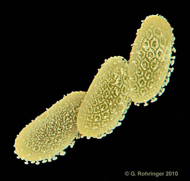
Dr. Gerhard Rohringer – Santa Barbara, California
Courtesy of Nikon Small World
Dr. John Runions – School of Life Sciences, Oxford Brookes University in Oxford, UK
Courtesy of Nikon Small World
Donald Schick – Pomona, California
Courtesy of Nikon Small World
Thomas Shearer – ClodStone Photography, LLC in Duluth, Minnesota
Courtesy of Nikon Small World
Dr. Peter Siver – Connecticut College in New London, Connecticut
Courtesy of Nikon Small World
Raymond Sloss – Northamptonshire Natural History Society in Northampton, UK
Courtesy of Nikon Small World
Craig Smith – Insight Photography in Fresno, California
Courtesy of Nikon Small World
Stanley Stein – Englewood, Colorado
Courtesy of Nikon Small World
Michael J. Stringer – I.D.R.S. Member (retired) in Westcliff-on-Sea, Essex, UK
Courtesy of Nikon Small World
Viktor Sykora – Institute of Pathophysiology, First Medical Faculty, Charles University in Prague, Czech Republic
Courtesy of Nikon Small World
Viktor Sykora – Institute of Pathophysiology, First Medical Faculty, Charles University in Prague, Czech Republic
Courtesy of Nikon Small World
Mayumi Wakazaki, Dr. Kiminori Toyooka – RIKEN Plant Science Center in Yokohama, Kanagawa, Japan
Courtesy of Nikon Small World
Wim van Egmond – Micropolitan Museum in Rotterdam, The Netherlands
Courtesy of Nikon Small World
Wim van Egmond – Micropolitan Museum in Rotterdam, The Netherlands
Courtesy of Nikon Small World
Alexa Vanegas – Universidad Nacional de Colombia in Medell\u00edn, Antioquia, Colombia
Courtesy of Nikon Small World
Bruno Vellutini – Centro de Biologia Marinha, Universidade de S\u00e3o Paulo in S\u00e3o Paulo, SP, Brazil
Courtesy of Nikon Small World
Bruno Vellutini – Centro de Biologia Marinha, Universidade de S\u00e3o Paulo in S\u00e3o Paulo, SP, Brazil
Courtesy of Nikon Small World
Philippe Verrees – Knokke, Belgium
Courtesy of Nikon Small World
David Walker – West Yorkshire, UK
Courtesy of Nikon Small World
Dr. Arlene Wechezak – Anacortes, Washington
Courtesy of Nikon Small World
Dr. Rong Wen – University of Miami, Bascom Palmer Eye Institute in Miami, Florida
Courtesy of Nikon Small World
Jim Wetzel – Presbyterian College in Clinton, South Carolina
Courtesy of Nikon Small World
Stephan Wolfsried – Waiblingen, Baden-W\u00fcrttemberg, Germany
Courtesy of Nikon Small World
Dr. Saiko Yoshida – University of Bern in Bern, Switzerland
Courtesy of Nikon Small World
Dr. Xiaowei Zhuang – Hughes Medical Institute, Department of Chemistry and Chemical Biology in Cambridge, Massachusetts
Courtesy of Nikon Small World

My name is Sonja Thompson. I've worked for TechRepublic since October of 1999, starting with the enewsletter team, then with the Premium Products group (creating books and CDs), as well as programming some of the elements on the site. After leading the Tech News team on TR, I jumped at the opportunity to switch gears and try my hand at video editing, podcasts, and other forms of multimedia on the site. I'm currently the host of the Smartphones blog, plus I edit the TR Dojo video series. \ \ I graduated from the University of Louisville. Since then, I've also completed several technology related courses from SmartPlanet. My goal is to learn about the TR community, interact with members on the site, and hopefully encourage more people to participate - and more often.


