Every year, Nikon hosts its Small World competition for microscopic scientific images. Here are the top 20 images from 2015, including photos of plants, animals, and their most interesting tiny bits.
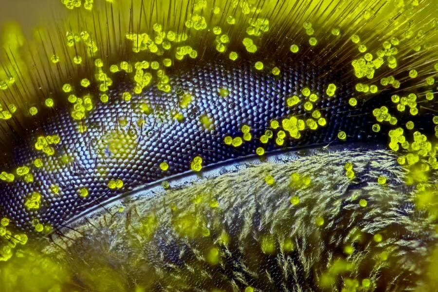
Hailing from Queensland, Australia, Grimm captured this image of a honey bee’s eye covered in dandelion pollen using reflected light.
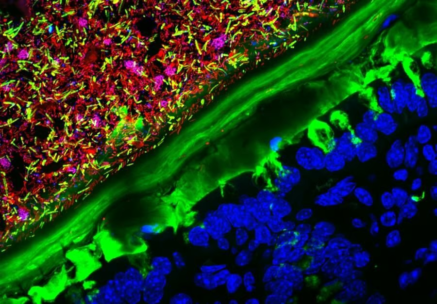
From the Stanford University School of Medicine, this image of a mouse colon colonized with human microbiota was captured using confocal microscopy.
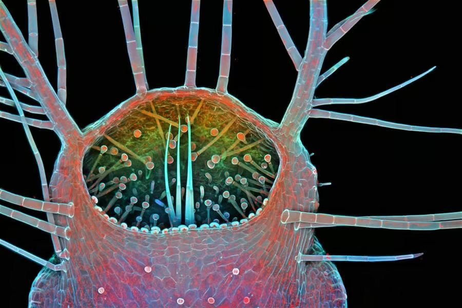
Also captured using confocal microscopy, this image shows the intake of a humped bladderwort plant. This freshwater carnivorous plant typically feeds on small invertebrates.
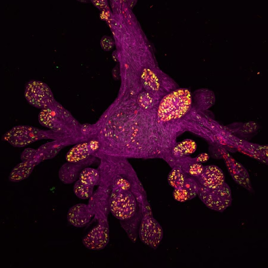
This human mammary gland organoid was actually grown in a lab.
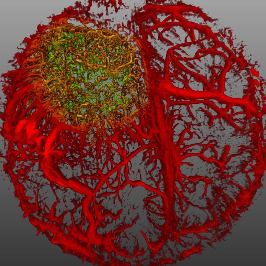
Here we have the vascular system of a mouse brain with glioblastoma, a malignant brain tumor.
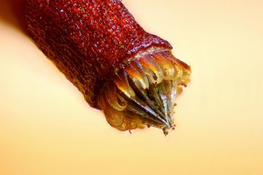
Using reflected light, Henri Koskinen of Helsinki, Finland captured this image of a spore capsule of a moss.
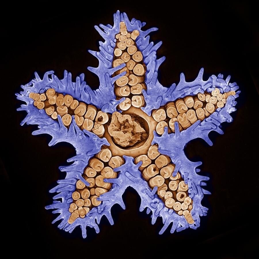
This starfish image was also captured using confocal microscopy, showing a fascinating amount of detail.
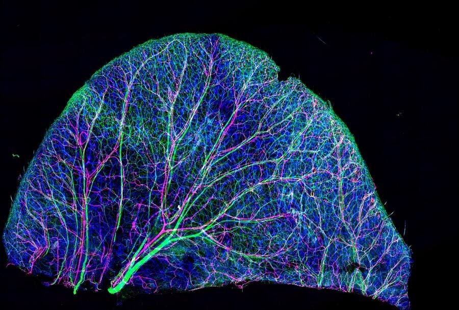
What you’re looking at is a mouse’s ear. Using microscopic imaging techniques, we are able to see the nerves and blood vessels in the skin of this mouse’s ear.
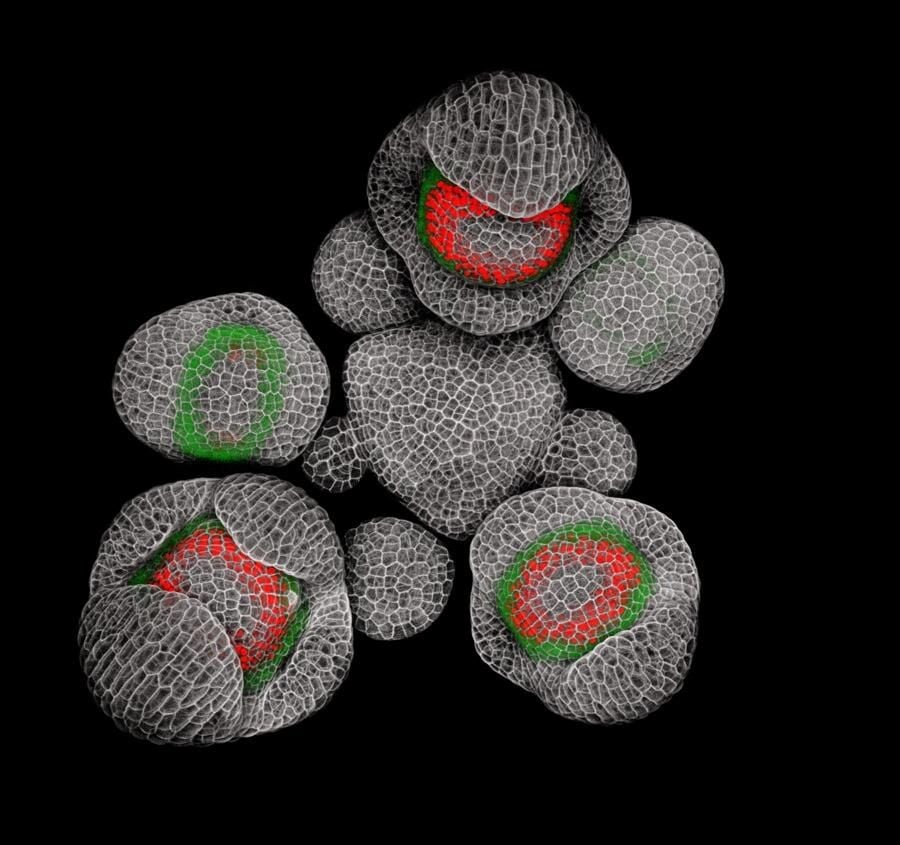
These are the buds of an Arabidopsis plant, sometimes referred to as rockcress.
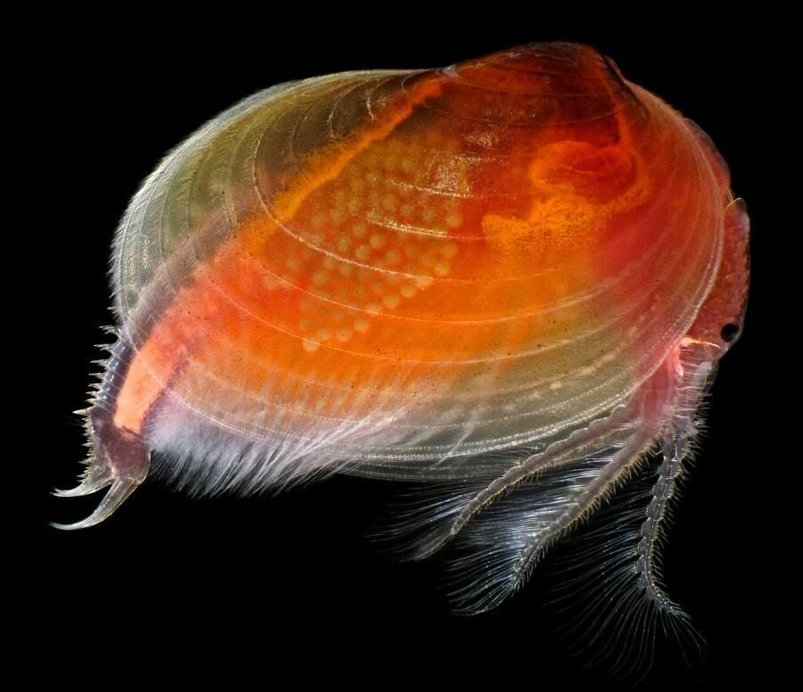
Clam shrimp are a type of crustacean, similar to the mollusc. This photo was taken in Calgary, Alberta, Canada.
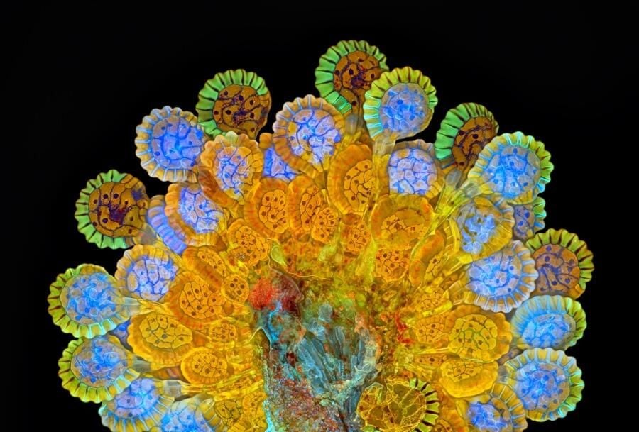
A sorus produces spores for a fern. Here we see it at different levels in the maturation process.
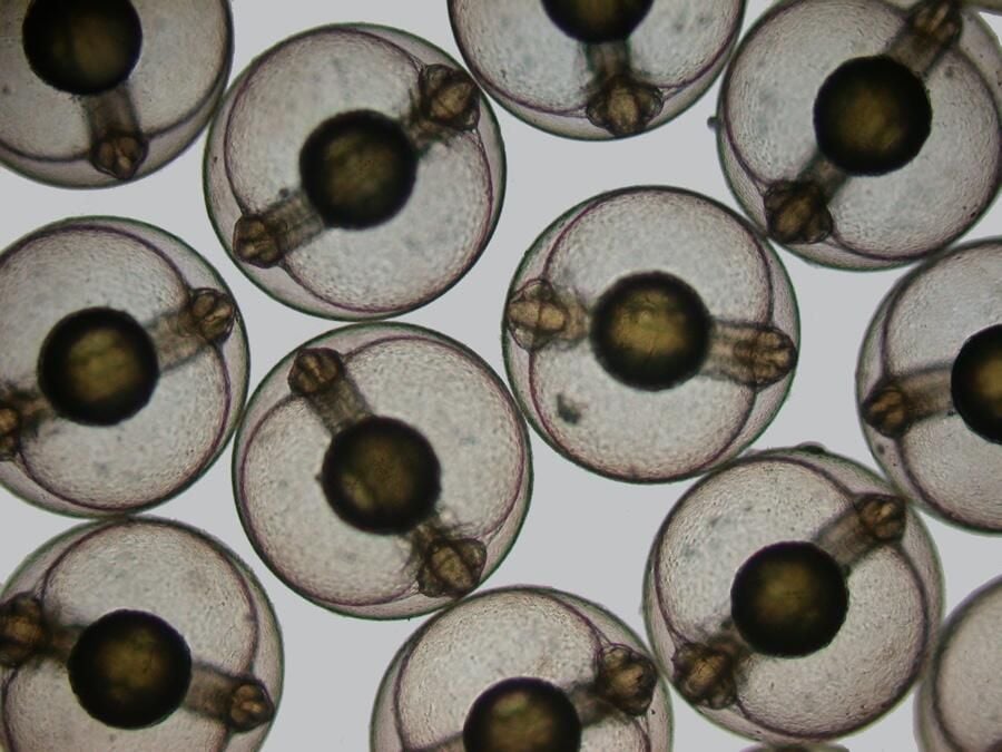
This image of developing sea mullet embryos was captured using brightfield imaging.
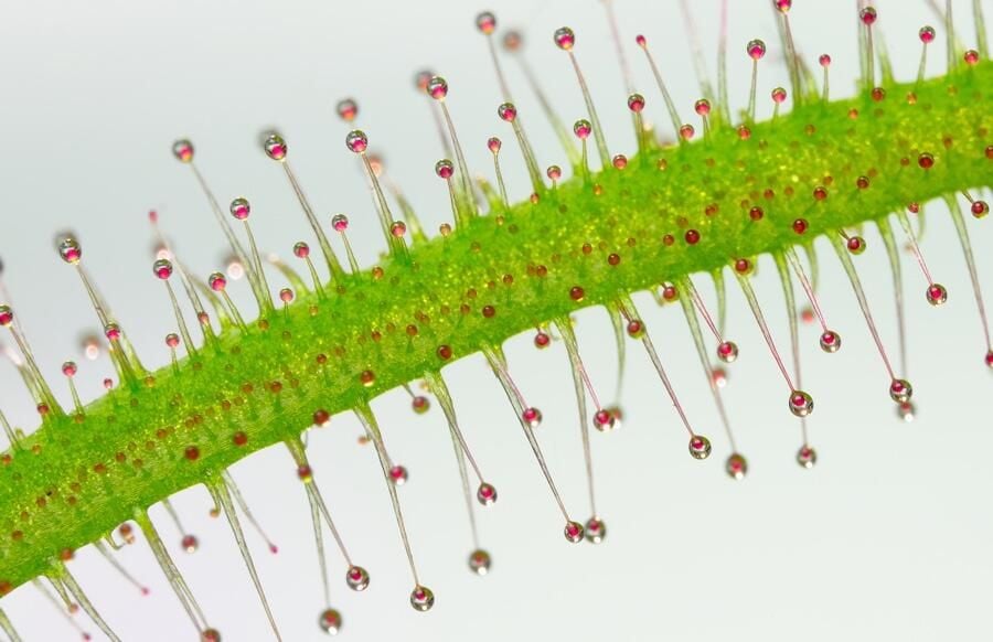
These tentacles belong to a plant in the Drosera, also known as sundew.
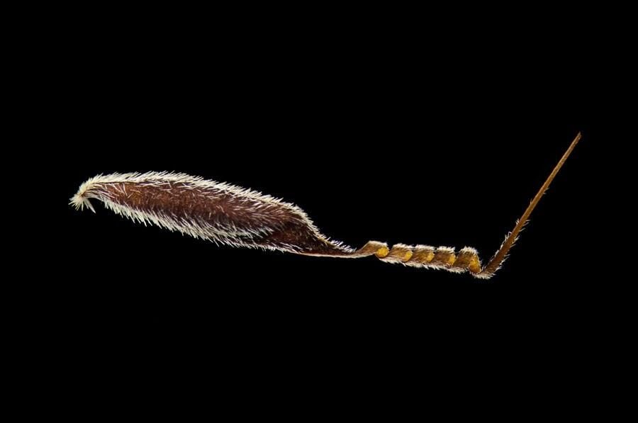
Sykora, of Prague, Czech Republic, took this image of Australian grass seed using dark field microscopy, which helps enhance the contrast.
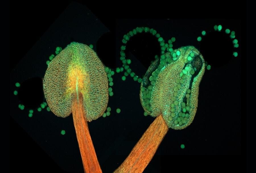
The anther is the part of a flower’s stamen that actually produces the pollen. This image shows the anther of a plant that is in the process of flowering.
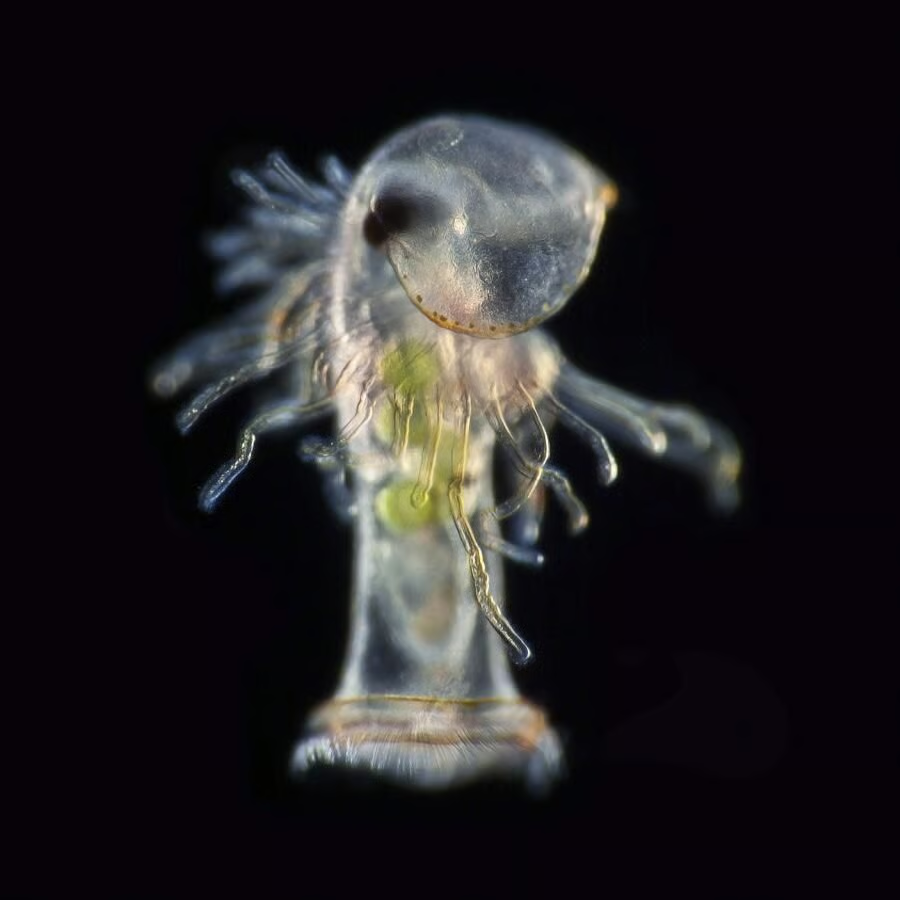
Rotifers, also known as wheel animals, are a form of pseudocoelomate animals.
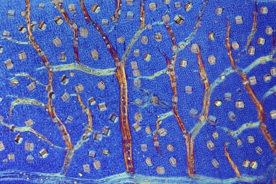
This image shows a black witch-hazel leaf producing crystals to defend itself against herbivores. It was captured using differential interference contrast, which helps to enhance the contrast of the sample.
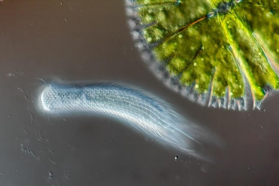
Switzerland’s Roland Gross caught this image of a hairyback, which is also known as a gastrotrich.
This image of a planktonic larva of a horseshoe worm was captured using dark field microscopy.
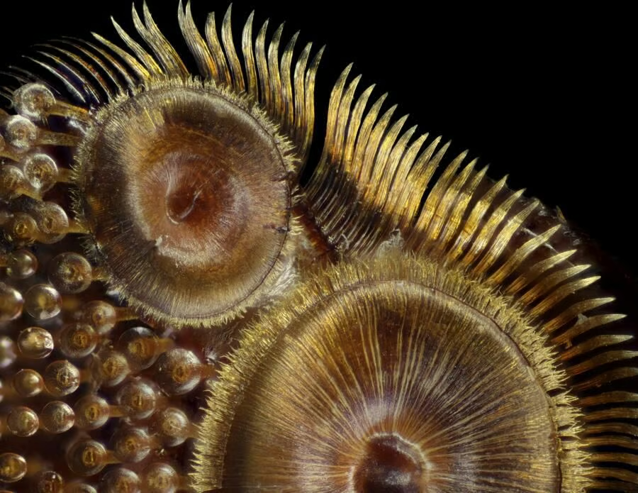
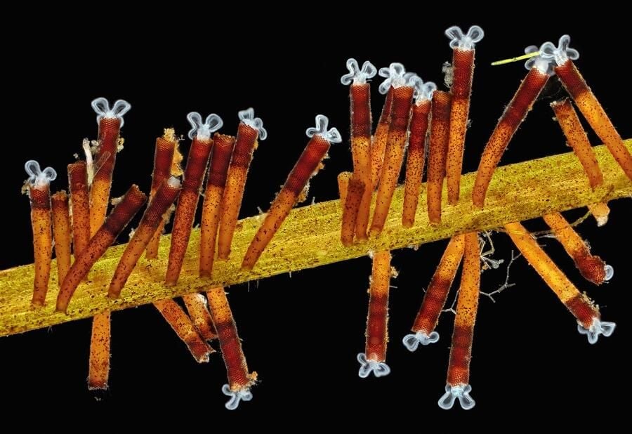
Here we see the suction cups on the foreleg of a diving beetle. On average, diving beetles are only 25 mm long.

Conner is a former Senior Editor for TechRepublic. He is now a Senior Research Analyst at 451 Research.


