How 3D bioprinting is changing the world: Photos of 10 great projects
Image 1 of 10

Human face
Human face
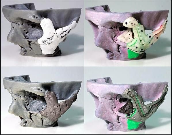
A man from Wales in the United Kingdom was in a motorcycle accident in 2012 and he has now received 3D printed implants on his face that successfully fixed injuries he sustained. The man broke his cheekbones, jaw, nose, and skull. CT scans were used to 3D print a symmetrical model of his skull, and then a printed titanium implant held the bones in place. The project was done by the Centre for Applied Reconstructive Technologies in Surgery.

Brain tissues
Brain tissues
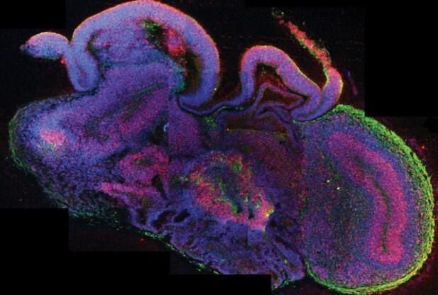
Scientists in Vienna, Austria printed brain tissues from stem cells. They call them u201ccerebral organoids.” The organoids grew to about four millimeters in size and could survive up to 10 months. Right now, the organoids are limited in their size because they cannot move around nutrients in the circulatory system. The researchers are using the tissue to study microcephaly, a condition where the brain stops growing. As with any other transplant, there is always the risk of rejection by the body with these 3D printed tissues.
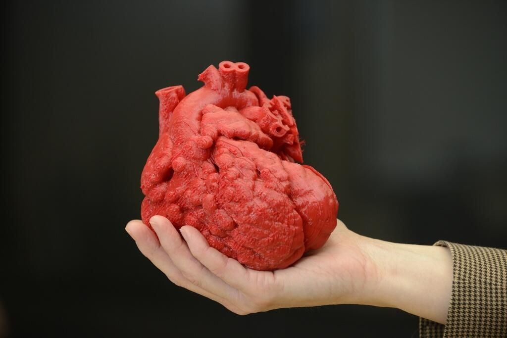
Human heart
Researchers at the University of Louisville in Louisville, Kentucky said they have successfully printed parts of a human heart using by printing with a combination of human fat cells and collagen. One liter of fat from someone can give them a huge number of cells that can be directly translated to patients. The team has not yet worked on putting any of the pieces of the heart together, but said the time to do so isn’t very far off.
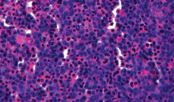
Liver tissue
In January, Organovo successfully printed samples of human liver tissue that were distributed to an outside laboratory for testing. The company is aiming for commercial sales later this year. The sets of 24 samples take about 30 minutes to produce. According to the company, the printed tissue responds to drugs similarly to a regular human liver.

Human skull
Human skull
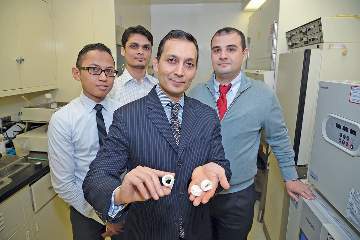
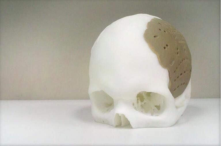
Oxford Performance Materials, a Connecticut-based company, recently received FDA approval for a 3D printed implant that replaced 75 percent of a man’s skull. The implant was not made from biological materials, but the material used is biocompatible and has a density similar to bones.
Blood vessels
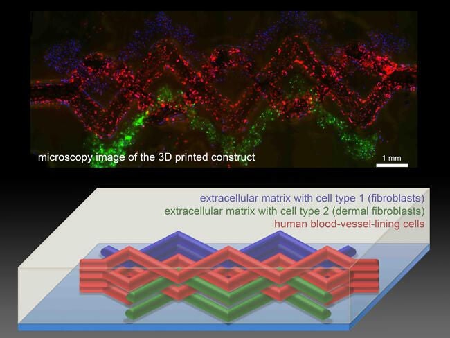
Harvard researchers said they have created tissue interlaced with blood vessels. The patch of tissue contained skin cells and biological structural materials interwoven with tiny blood vessels. This is the first 3D printed structure that provides a major step in making tissues that are realistic enough to test drug safety and effectiveness.

Pancreatic endocrine tissues
Pancreatic endocrine tissues
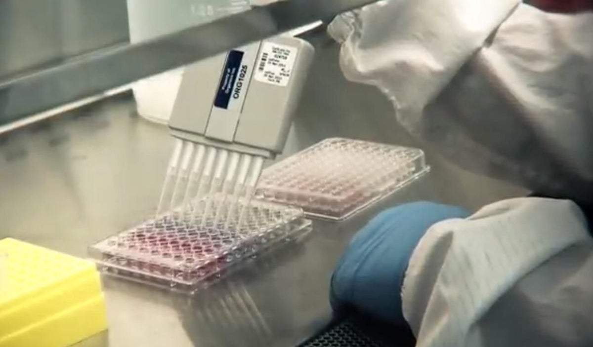
A University of Iowa engineering professor received an award from the National Science Foundation to 3D print pancreatic endocrine tissues. The cells are taken from an individual’s body. The professor said it will have an impact on the ability to print glucose-sensitive tissues as an alternative solution to effectively manage diabetes.

Eyes
Eyes
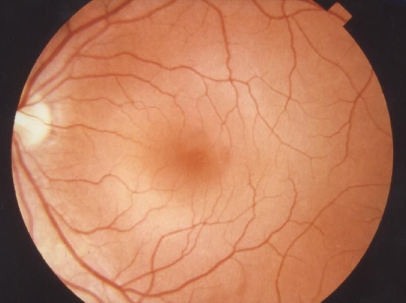
At the end of last year, researchers in the UK used an inkjet printer to 3D print living retinal cells of adult rats, which could be used in the future as replacement eye tissues. The researchers said it may aid in the cure for blindness, specifically for glaucoma and macular degeneration, and is a very important step for regenerative medicine. This study was the first of its kind to show that the cells can be stacked on top of one another without being damaged. They were not distorted when printed out at such a high speed.
Disclaimer: We’re still trying to understand how exactly this would work, and we haven’t yet found a good medical source that clearly explains it.
Trachea
Researchers at Mount Sinai built a trachea with a 3D printer and biological membrane. The membrane was seeded with a stem cell solution. The cells were transformed into cartilage precursors and implanted into a baby pig. After two months, the pig has had no major issues with the implant. Because the cells used for this would be from the person’s body, it should be well-tolerated.
Skin
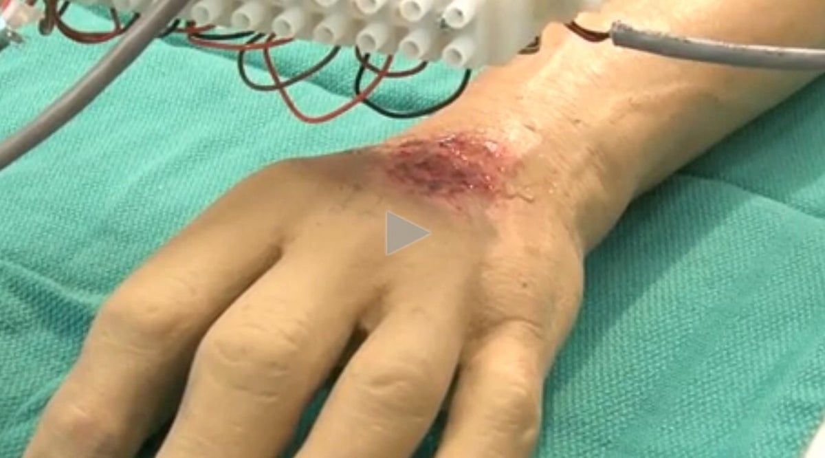
Scientists at Wake Forest School of Medicine designed a printer that can directly print skin cells onto burn wounds. The traditional treatment for severe burns is to cover them with healthy skin harvested from another part of the body, but often times there isn’t enough. With this new machine, a scanner determines the size and depth of the skin, and layers the appropriate number of cells on the wound. Doctors only need a patch of skin one-tenth of the size of the wound to grow enough for this process.
-
-
Account Information
Contact Lyndsey Gilpin
- |
- See all of Lyndsey's content
